Biomedical Illustrator Medical & Biological Illustrations Laurie O

Leaf Structure photo Botany, Teaching biology, Biology
Structure Types Modification Function Key Points Let's learn more about the morphology of leaves, parts of a leaf, different types of leaves and their modifications. Structure of a Leaf Leaves are thin, flat organs responsible for photosynthesis in the plants. It develops laterally at the node.
Structure of a leaf
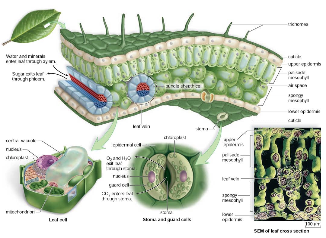
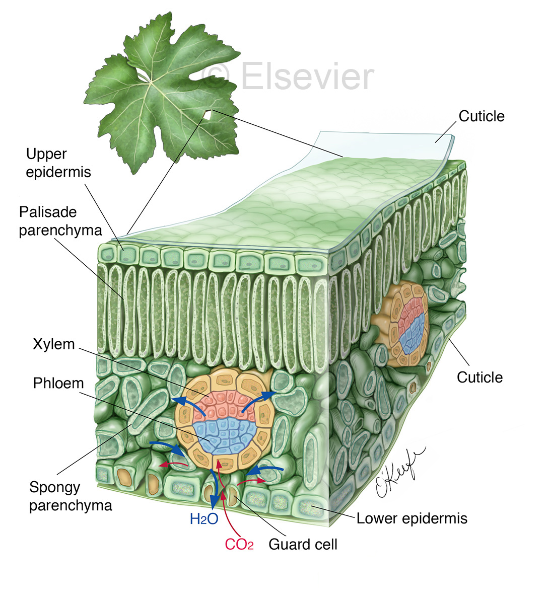
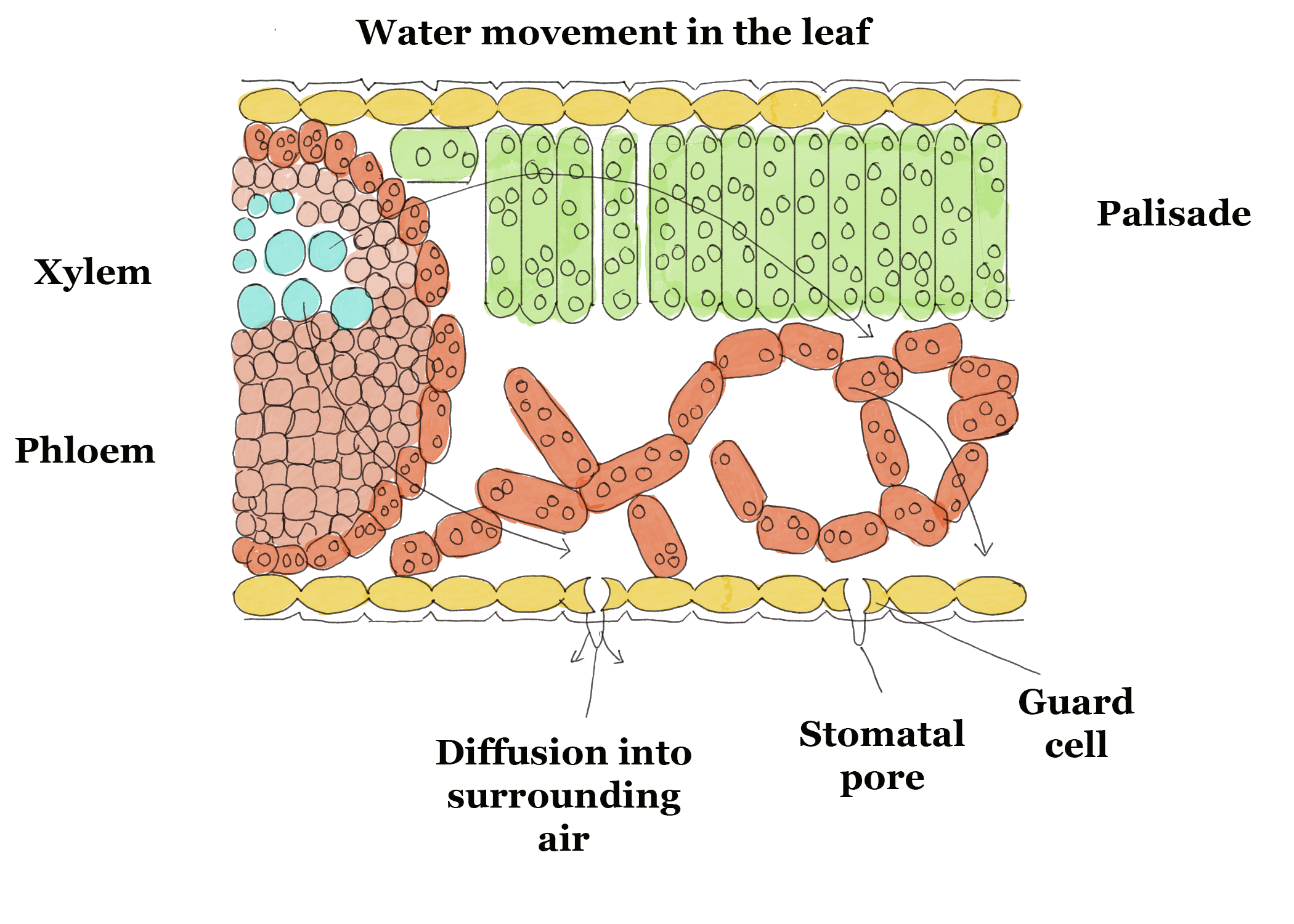
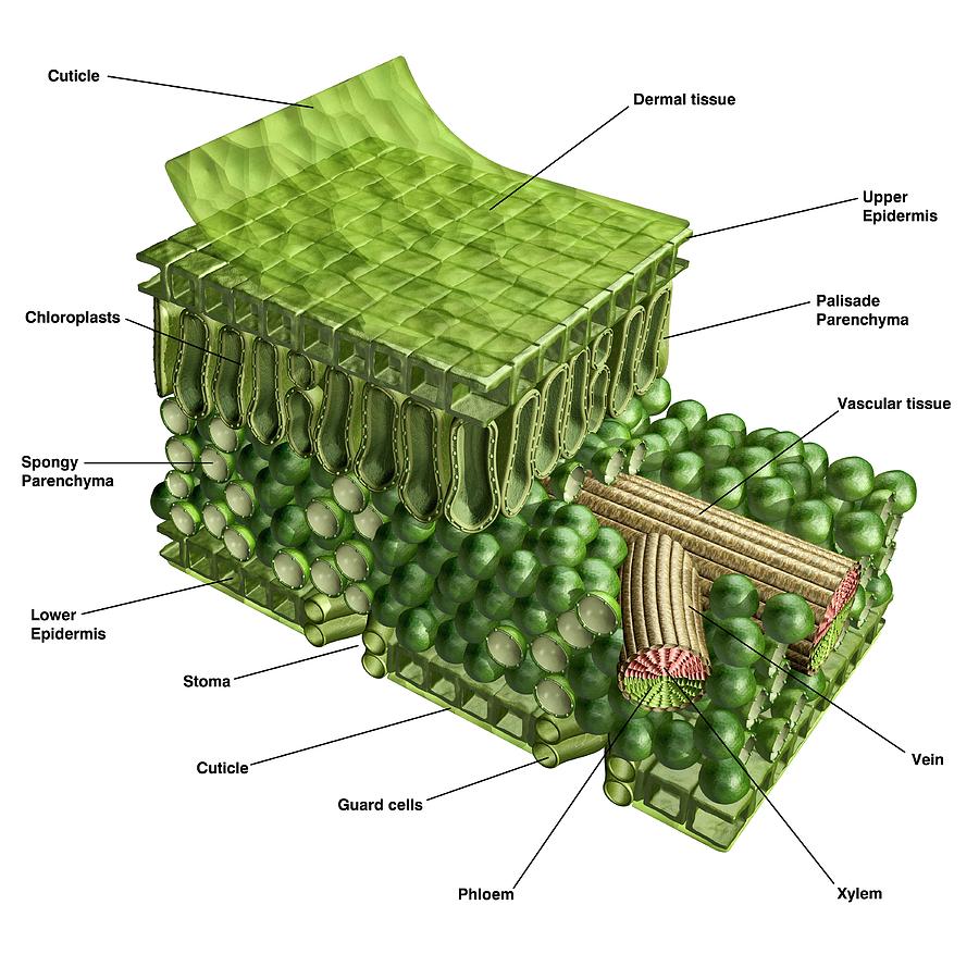
Like the stem, the leaf contains vascular bundles composed of xylem and phloem (Figure 3.4.2.6 − 7 3.4.2. 6 − 7 ). When a typical stem vascular bundle (which has xylem internal to the phloem) enters the leaf, xylem usually faces upwards, whereas phloem faces downwards. The conducting cells of the xylem (tracheids and vessel elements.
Biomedical Illustrator Medical & Biological Illustrations Laurie O
They are attached by a continuous vascular system to the rest of the plant so that free exchange of nutrients, water, and end products of photosynthesis (oxygen and carbohydrates in particular) can be carried to its various parts. Leaves are initiated in the apical bud (growing tip of a stem) along with the tissues of the stem itself.
/Leaf_Tissue_Structure-56e6d04c5f9b5854a9f9494e.jpg)
Anatomy of a Leaf, Use These Leaf Parts to Identify a Tree
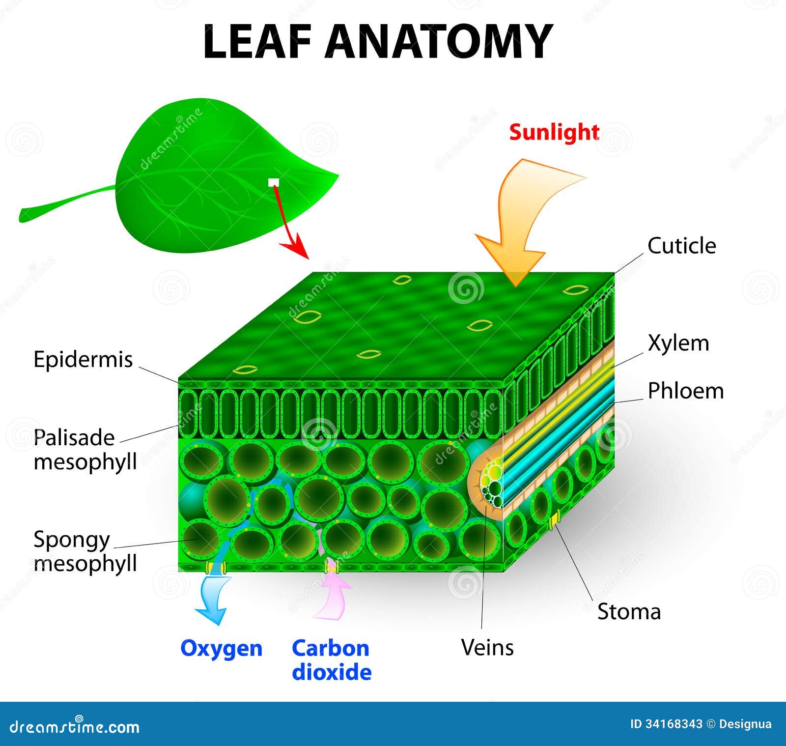
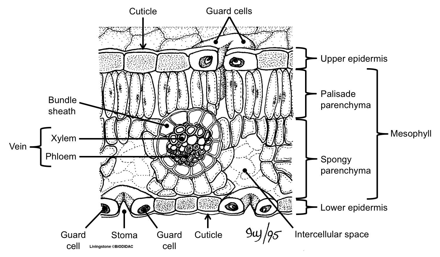
The air space found between the spongy parenchyma cells allows gaseous exchange between the leaf and the outside atmosphere through the stomata. In aquatic plants, the intercellular spaces in the spongy parenchyma help the leaf float. Both layers of the mesophyll contain many chloroplasts. Figure 30.10. 1: Mesophyll: (a) (top) The central.
Leaf Structure, Types, Functions GCSE Biology Revision
Leaf parts and directional terms. Left: Diagram of a simple leaf showing the basic parts, including the petiole (stalk), lamina (blade), veins (strands of vascular tissue), margin (edge of the lamina), apex of the lamina, and base of the lamina.Right: Diagram of a leaf attached to a stem showing terms for directionality: adaxial (upper leaf surface), abaxial (lower leaf surface), proximal.
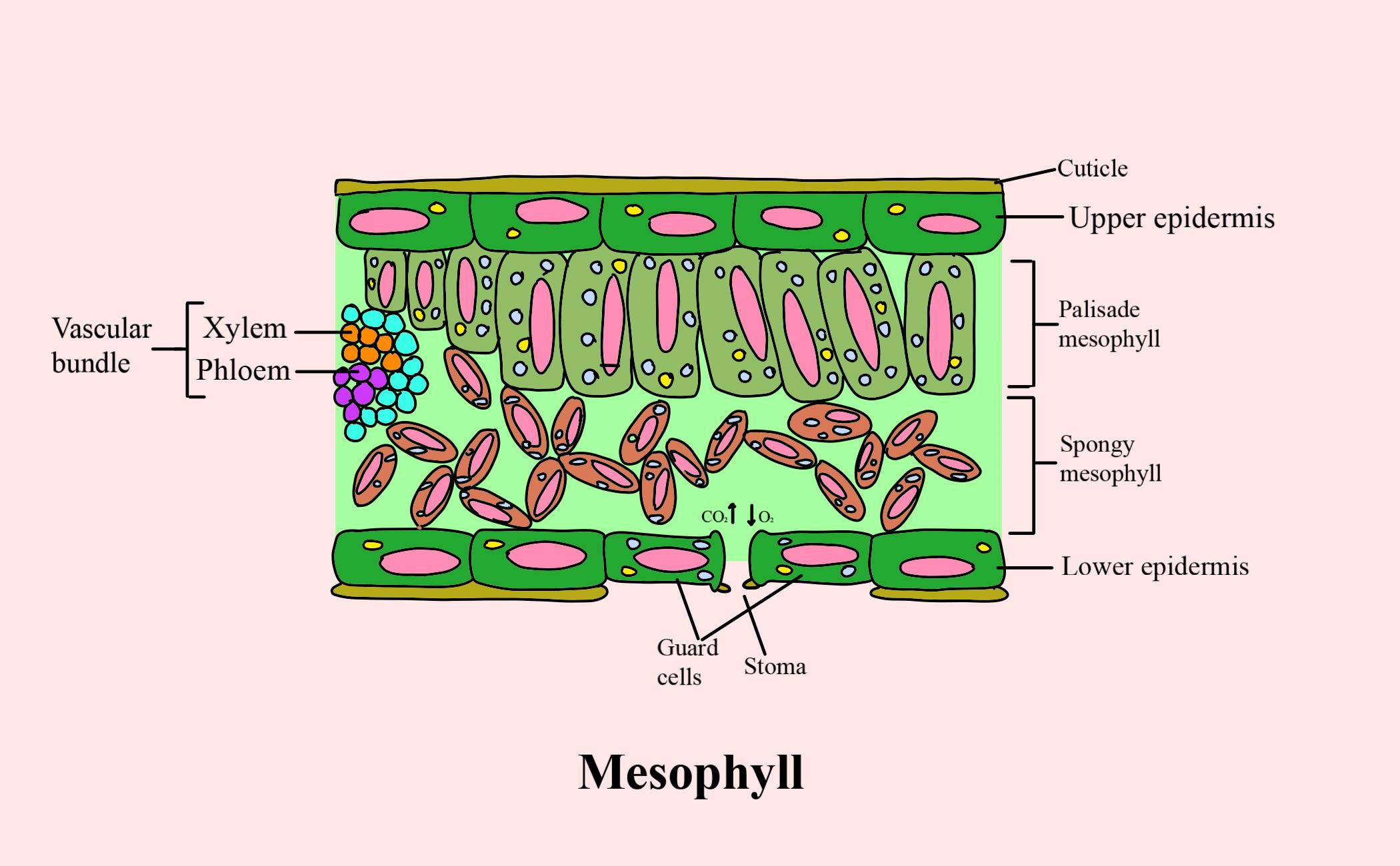
The mesophyll of leaf consists of (a) Spongy parenchyma cells (b
Internal Structure of Leaf (With Diagram) Article Shared by ADVERTISEMENTS: In this article, we propose to discuss about the internal structure of leaf. The foliage leaves are characterised by green colour, thinness and flatness. They develop as protrusions from the shoot apex and are organs of limited growth.
Plant Leaf Structure Photograph by Carlos Clarivan Pixels
Features of leaves and their functions The role of stomata The control gas exchange in the leaf. Each stoma can be open or closed, depending on how its guard cells are. The stomata can open and.

Parts of a leaf diagram. Preschool & PreK Science/Sensory
Overview By the end of this section, you will be able to do the following: Identify the parts of a typical leaf Describe the internal structure and function of a leaf Compare and contrast simple leaves and compound leaves List and describe examples of modified leaves
Leaf anatomy stock illustration. Illustration of lower 34168343
Parts of a Leaf Diagram 1. Petiole It is the stalk that connects a leaf to the stem of the plant, it is made of complex conducting tissues called vascular tissues. Functions Providing support to the leaf and keeps it erect Transporting water and nutrients absorbed by the roots to the leaves
:max_bytes(150000):strip_icc()/parts_of_a_leaf-56abaed23df78cf772b5625a.jpg)
Plant Leaves and Leaf Anatomy
Structure of a Leaf (With Diagram) | Plant Organs | Biology Article Shared by ADVERTISEMENTS: In this article we will discuss about the structure of a leaf with the help of a diagram. A leaf is a compromise between two conflicting evolutionary pressures.

Leaf structure narrated YouTube
2. Isobilateral Leaf: This cannot be differentiated internally into two regions. The structure on both the surfaces is quite similar (isos = equal; bi = two; lateris = side). It is also called a unifacial leaf or isolateral leaf. The stomata in this case are usually present in both the epidermal layers and thus, it may be known as amphistomatic.

gleeson11biology / C6
Figure 30.8.1 30.8. 1: Parts of a leaf: A leaf may seem simple in appearance, but it is a highly-efficient structure. Petioles, stipules, veins, and a midrib are all essential structures of a leaf. Within each leaf, the vascular tissue forms veins. The arrangement of veins in a leaf is called the venation pattern.

Leaf anatomy vector illustration diagram Anatomy, Medical
The structure of the umbrella tree leaf is typical of leaves in general (Above left photo). It has an outer layer, the epidermis, which produces a waxy waterproof coating. The epidermis of the undersurface produces guard cells, which swell and shrink to close and open the pores (stomata) which control the loss of water vapor (transpiration) and.
Leaf Structure & Evolution Digital Atlas of Ancient Life
The photo on the left is a palmate leaf, the diagram on the right is a pinnate leaf. Photo by Maria Morrow, CC-BY 4.0. Diagram on the right from Chapter 3.4.2, Botany, by Algiers, Ha, and Morrow. The Leaf Blade and Vascular Arrangement. The leaf blade is (usually) the flat, photosynthetic part of the blade. In eudicots, the leaf will have a.
leaf structure Labelled diagram
Leaves are the powerhouse of plants. In most plants, leaves are the major site of food production for the plant. Structures within a leaf convert the energy in sunlight into chemical energy that the plant can use as food. Chlorophyll is the molecule in leaves that uses the energy in sunlight to turn water (H 2 O) and carbon dioxide gas (CO 2.

Labeled Diagram Of A Leaf hubpages
How do they work? An microphotograph of a stoma shows the two guard cells which regulate its opening and closure to limit water loss, excrete oxygen, and absorb carbon dioxide. The openings or pores in stomata are formed by two specialized sclerenchymal cells, the guard cells ( Figure above ).
