Anatomy of brain labeled diagram Science
The Brain Scientific Publishing
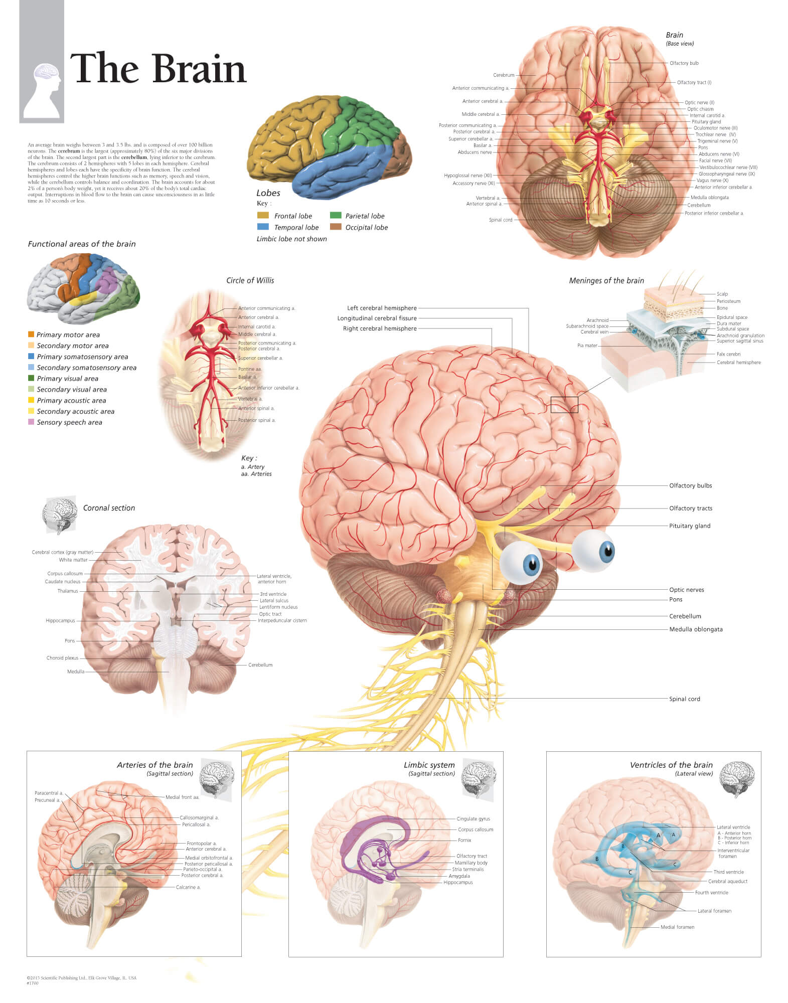
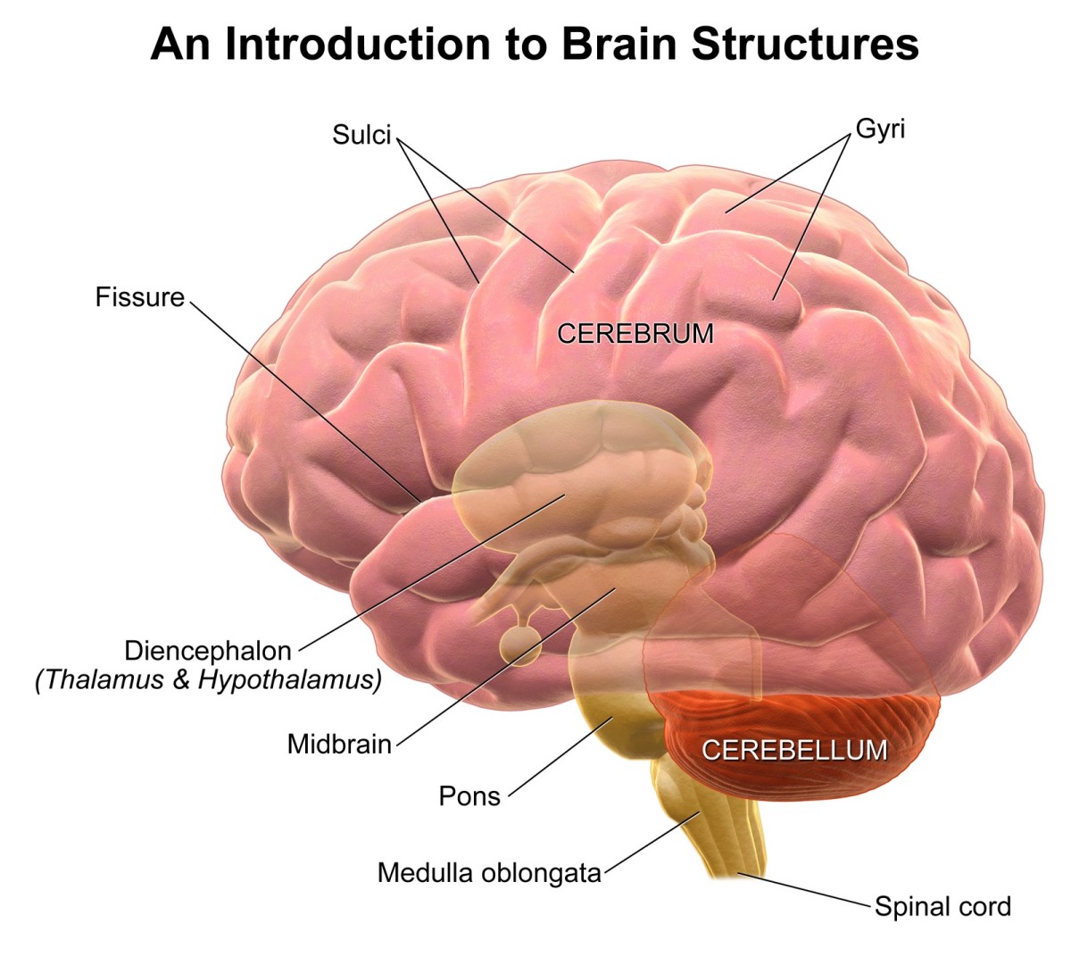
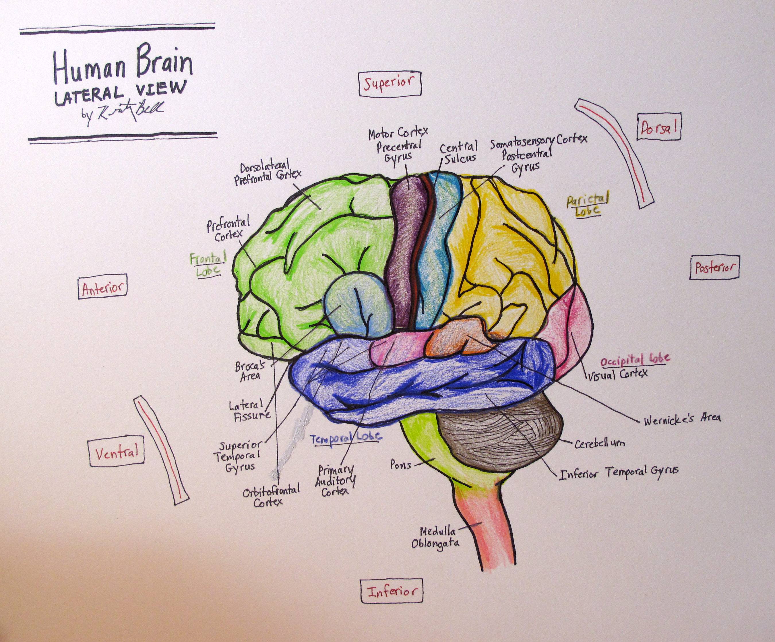
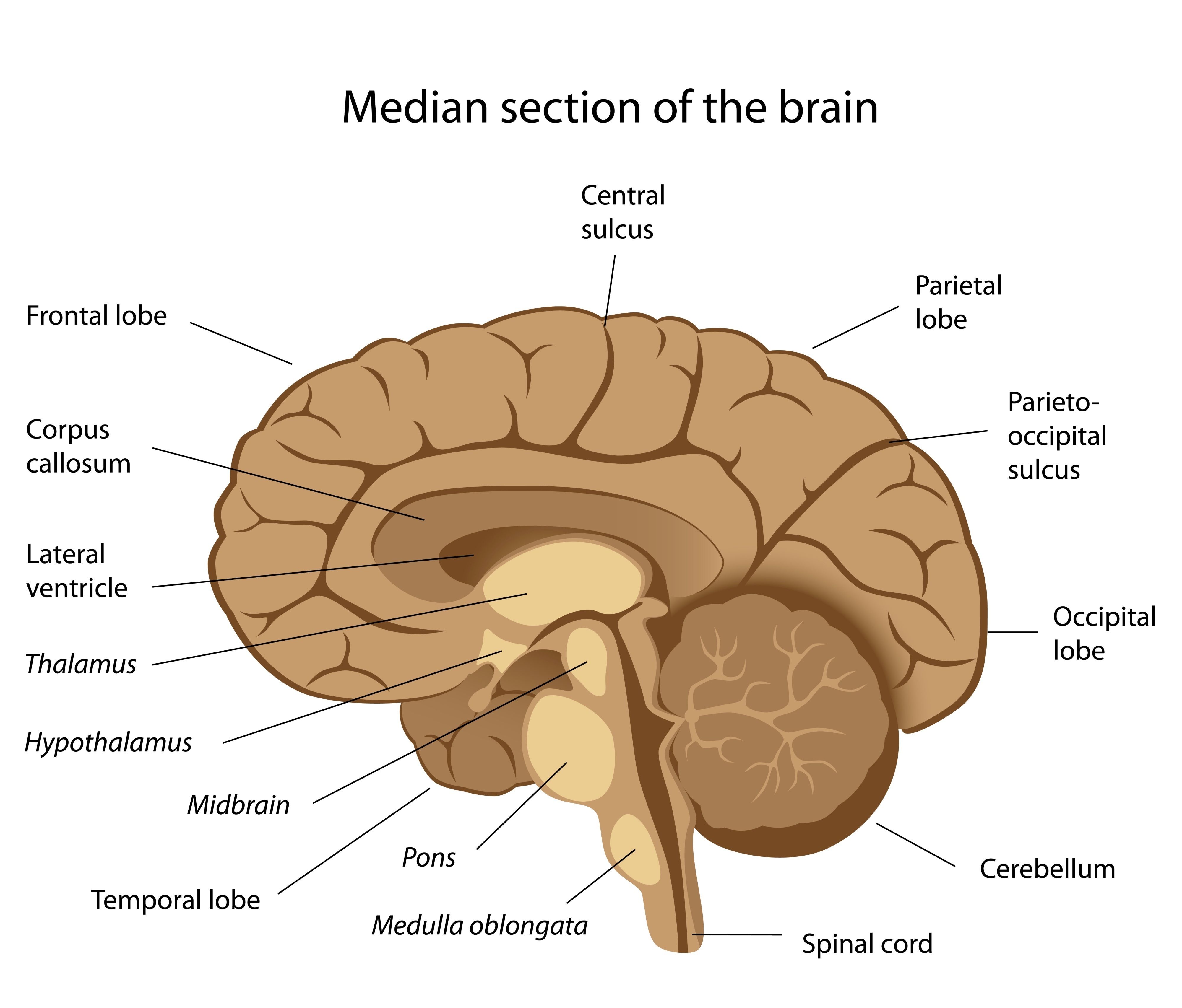
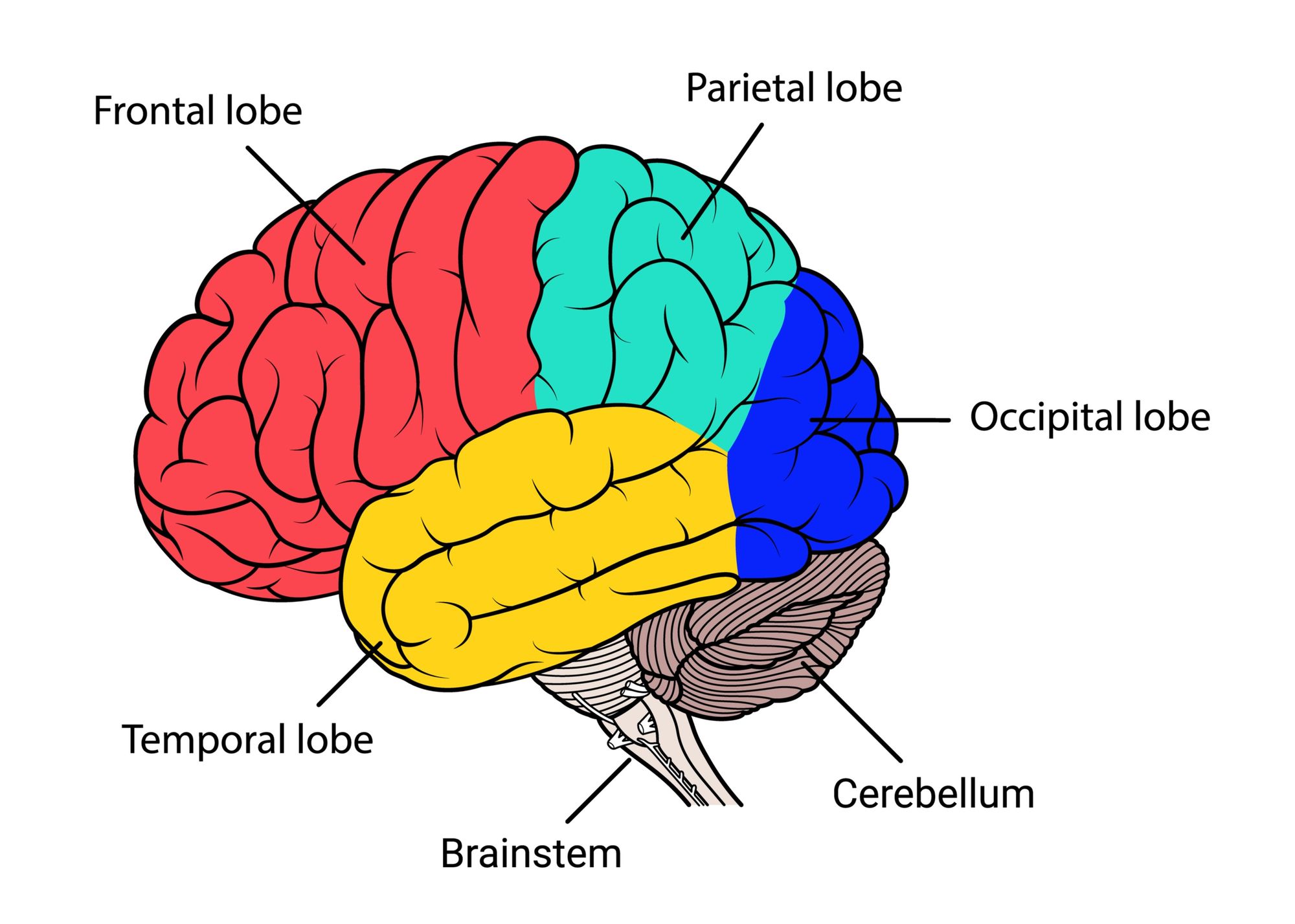
Brain Parts The brain is composed of the cerebrum, cerebellum, and brainstem (Fig. 1). Figure 1. The brain has three main parts: the cerebrum, cerebellum, and brainstem. Cerebrum The cerebrum is the largest and most recognizable part of the brain. It consists of grey matter (the cerebral cortex ) and white matter at the center.
The Human Brain Facts, Anatomy, and Functions HubPages
Exploring the human brain (Image credit: Albert L. Rhoton Jr., MD, 2007.) Dr. Albert Rhoton of the University of Florida has collected an unparalleled library of brain-anatomy images..

Image result for labeled diagram of the brain Brain diagram, Human
Diagrams Diagrams are the perfect way to get orientated with a structure's detailed anatomy. Read on to see how we recommend using them. If you need some help with labeling the following diagrams, check out this video where we show you how to do it step-by-step: Labeled brain diagram
Brain Jack Image กรกฎาคม 2013
The brain directs our body's internal functions. It also integrates sensory impulses and information to form perceptions, thoughts, and memories. The brain gives us self-awareness and the ability to speak and move in the world. Its four major regions make this possible: The cerebrum, with its cerebral cortex, gives us conscious control of our.
Drawing Of The Brain With Labels at GetDrawings Free download
The human brain is the central organ of the human nervous system, and with the spinal cord makes up the central nervous system.The brain consists of the cerebrum, the brainstem and the cerebellum.It controls most of the activities of the body, processing, integrating, and coordinating the information it receives from the sense organs, and making decisions as to the instructions sent to the.
.jpg)
Picture of human Brain Human Anatomy
The Anatomy of the Human Brain: 3D Model The Human Brain By: Tim Taylor Last Updated: Jul 30, 2020 2D Interactive NEW 3D Rotate and Zoom Anatomy Explorer HINDBRAIN AND MIDBRAIN Brain Stem Inferior Colliculus Medulla Oblongata Pons Quadrigeminal Lamina Superior Colliculus Cerebellum Cerebellar Peduncle 4th Ventricle Cerebral Aqueduct Choroid Plexus
Free Brain Diagram, Download Free Brain Diagram png images, Free
Anatomy of the brain: how to view anatomical labels. This module is a comprehensive and affordable learning tool for medical students and residents and especially for neuroradiologists and radiation oncologists. It provides access to an atlas and to images in axial planes, allowing the user to learn and review neuroanatomy interactively. Images.

educational reflections with Mr. P, OCT (formerly Rumblings from 52
We introduce the Mindboggle-101 dataset, the largest and most complete set of free, publicly accessible, manually labeled human brain images. To manually label the macroscopic anatomy in magnetic resonance images of 101 healthy participants, we created a new cortical labeling protocol that relies on robust anatomical landmarks and minimal manual edits after initialization with automated labels.

Alila Medical Media Neurology Images & Videos
Illustration Picture of Brain Anatomy - Brain Picture of Brain The brain is the complex organ responsible for processing sensory information (sound, touch, taste, sight, and smell). The brain controls voluntary and involuntary movements. Signals from the brain tell muscles to contract.

Francisco's AP Macroeconomics Blog Psychology Unit 4Biological Basis
Healthy brain Summary The brain connects to the spine and is part of the central nervous system (CNS). The various parts of the brain are responsible for personality, movement, breathing, and.
Anatomy of brain labeled diagram Science
3D Brain. This interactive brain model is powered by the Wellcome Trust and developed by Matt Wimsatt and Jack Simpson; reviewed by John Morrison, Patrick Hof, and Edward Lein. Structure descriptions were written by Levi Gadye and Alexis Wnuk and Jane Roskams.
Label the Brain
3,226 labeled brain anatomy stock photos, 3D objects, vectors, and illustrations are available royalty-free. See labeled brain anatomy stock video clips Filters All images Photos Vectors Illustrations 3D Objects Sort by Popular Somatic vs autonomic nervous system division in human brain outline diagram.
:max_bytes(150000):strip_icc()/human-brain-regions--illustration-713784787-5973a8a8d963ac00103468ba.jpg)
Label Parts Of The Brain Pensandpieces
Relating structure and function in the human brain: relative contributions of anatomy, stationary dynamics, and non-stationarities. PLoS Computational Biology 10 (3):e1003530. doi:10.1371/journal.
PostStroke Dizziness How Vestibular Therapy Can Help
How does the brain work? The brain sends and receives chemical and electrical signals throughout the body. Different signals control different processes, and your brain interprets each. Some make you feel tired, for example, while others make you feel pain.

Labeled Pictures Of The Brain koibana.info Brain anatomy, Brain
Anatomy of the brain (MRI) - cross-sectional atlas of human anatomy. The module on the anatomy of the brain based on MRI with axial slices was redesigned, having received multiple requests from users for coronal and sagittal slices. The elaboration of this new module, its labeling of more than 524 structures on 379 MRI images in three different.
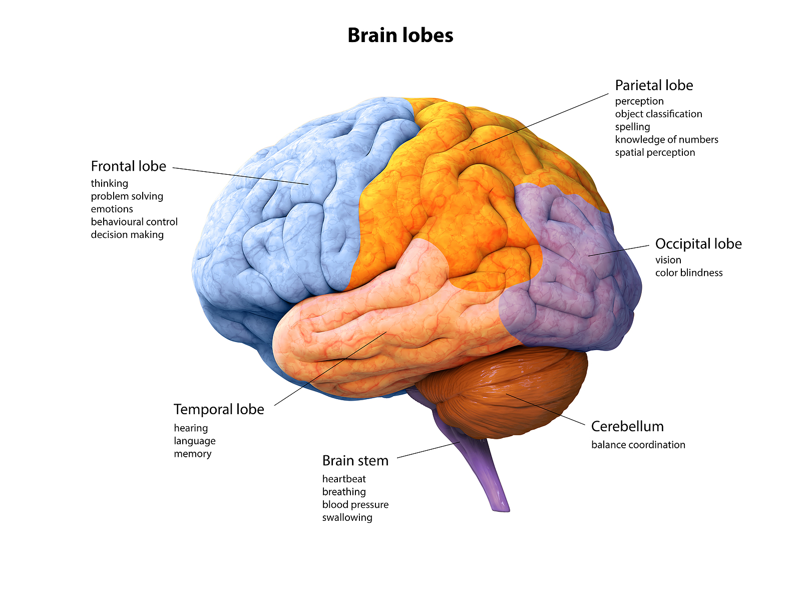
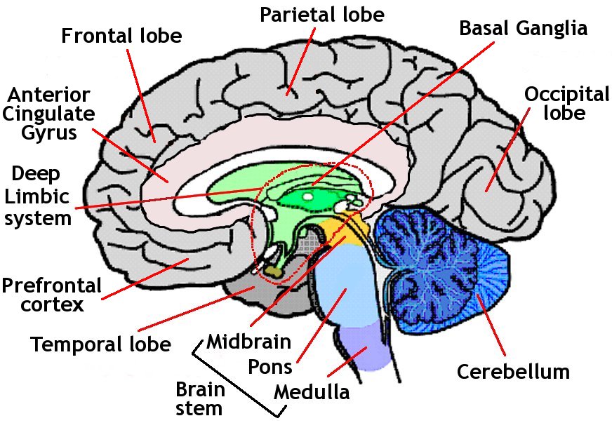
25 Facts about the lobes of the brain MooMooMath and Science
Brain Basics: Know Your Brain The brain is the most complex part of the human body. This three-pound organ is the seat of intelligence, interpreter of the senses, initiator of body movement, and controller of behavior. Lying in its bony shell and washed by protective fluid, the brain is the source of all the qualities that define our humanity.
