Human Skin Cell Under Microscope Micropedia Images and Photos finder

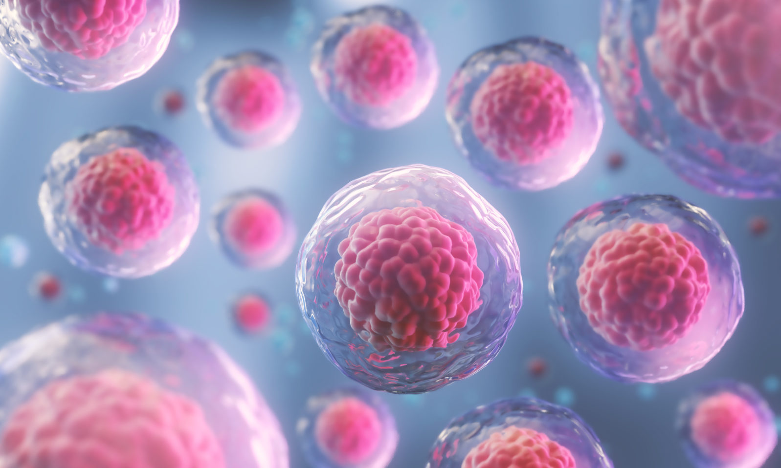
Premium AI Image Human cell microscope
In Figure 3.1.2 3.1. 2, only one edge of the tissue slice has epithelial cells. In Figure 3.1.2 3.1. 2 A that edge is indicated with an arrow, but when looking at a specimen under a microscope, you have to figure out for yourself where the edge with the epithelial cells is. Figure 3.1.2 3.1. 2: A slice of a trachea.
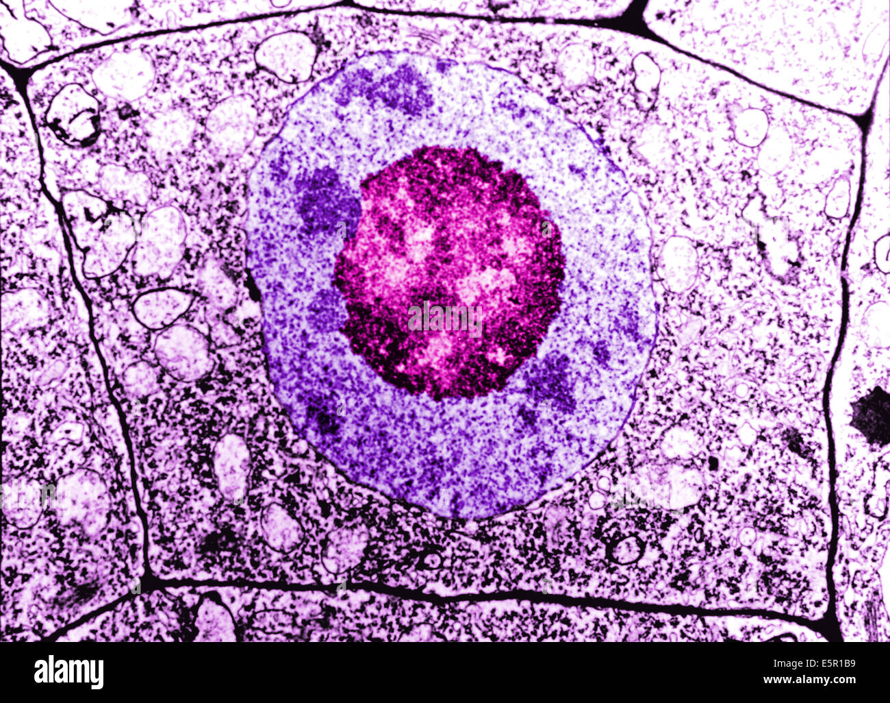
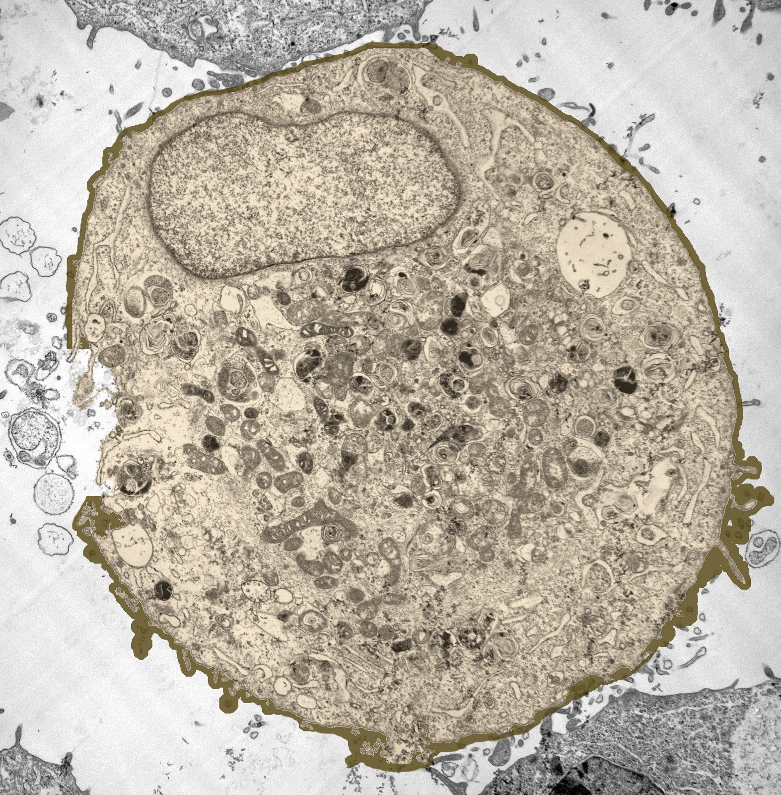
Electron Microscopy of a normal human cell, The cell membrane, nucleus
This includes human cells and many other types of cells that you will be studying in this class. The microscope you will be using uses visible light and two sets of lenses to produce a magnified image.. Biologists typically use microscopes to view all types of cells, including plant cells, animal cells, protozoa, algae, fungi, and bacteria.
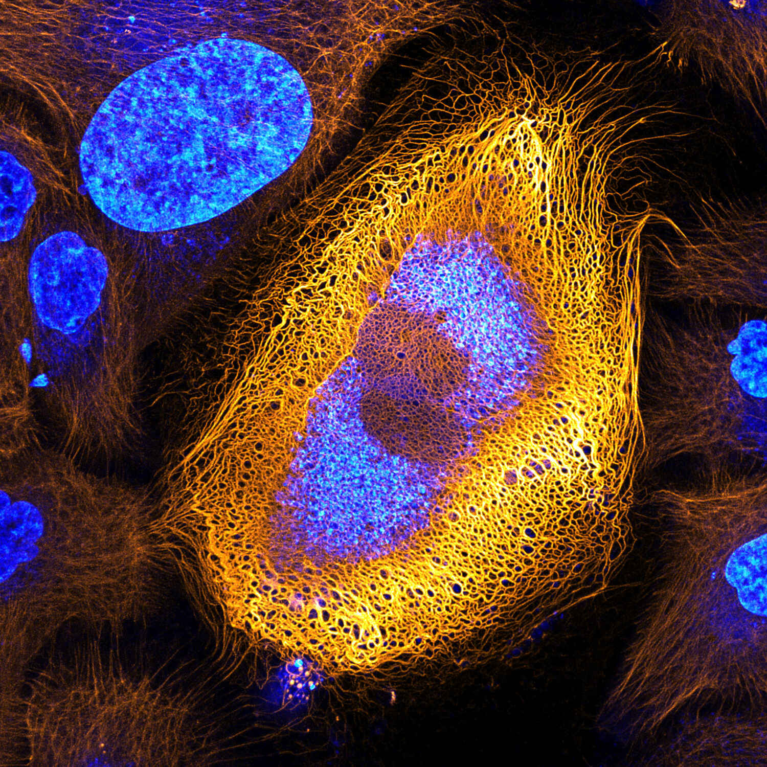
Stunning Microscopic View of Human Skin Cells Wins 2017 Nikon Small
The optical microscope is a useful tool for observing cell culture. However, successful application of microscope observation for culture evaluation is often limited by the skill of the operator and/or the lower reproducibility of visual evaluations. Automatic imaging and analysis for cell culture evaluation helps address these issues, and is seeing more and more practical use.

Human Animal Cell Under Microscope. Stock Illustration Illustration
A microscope is an instrument that magnifies objects otherwise too small to be seen, producing an image in which the object appears larger. Most photographs of cells are taken using a microscope, and these pictures can also be called micrographs. From the definition above, it might sound like a microscope is just a kind of magnifying glass.
4.2 Discovery of Cells and Cell Theory Human Biology
When dividing, they look like short, rod-like, tightly coiled structures and now called The human cells typically contain 46 chromosomes (except mature sex cells which contain a haploid number of chromosomes, i.e., 23 chromosomes). The DNA molecules carry the master code for making all of the enzymes and other proteins of a cell.

blood cells, cells, human, electron microscope, scan, blood
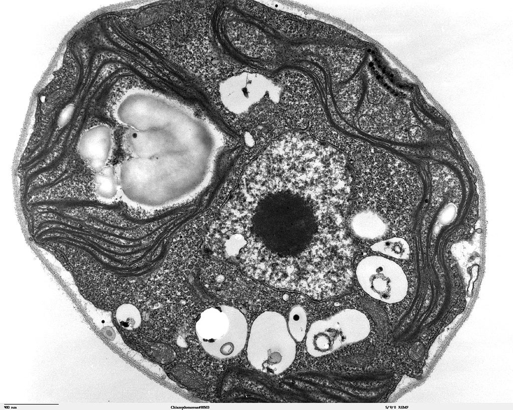
Looking at the Structure of Cells in the Microscope - Molecular Biology of the Cell - NCBI Bookshelf A typical animal cell is 10-20 μm in diameter, which is about one-fifth the size of the smallest particle visible to the naked eye.

Premium AI Image Human cell microscope
the cell structure under the microscope. cell, the waves are still "in phase"; this is no longer the case once they have passed through the various cell components. It is not possible for the human eye to rec-ognize these phase shifts. It can only distinguish between different intensities and colors. The phase contrast method

Human Cells Under Microscope HighRes Stock Photo Getty Images
Light Microscopes. To give you a sense of cell size, a typical human red blood cell is about eight millionths of a meter or eight micrometers (abbreviated as eight μm) in diameter; the head of a pin of is about two thousandths of a meter (two mm) in diameter. That means about 250 red blood cells could fit on the head of a pin.
Are the Brain Cells in a Dish That Learned Pong Conscious? Mind Matters
Muscle tissue is made up of cells that have the unique ability to contract or become shorter. There are three major types of muscle tissue, as pictured in Figure 5.3.14 5.3. 14: skeletal, smooth, and cardiac muscle tissues. Skeletal muscles are striated, or striped in appearance, because of their internal structure.
Human Skin Cell Under Microscope Micropedia Images and Photos finder
Cheek Cells Under The Microscope Sci- Inspi 334K subscribers Subscribe Subscribed 914K views 6 years ago Human cheek cells are made of simple squamous epithelial cells, which are flat.

Electron microscope, Microscopy, Scanning electron microscope
Observing human cheek cells under a microscope is a simple way to quickly view and learn about human cell structure. Many educational facilities use the procedure as an experiment for students to explore the principles of microscopy and the identification of cells, and viewing cheek cells is one of the most common school experiments used to teach students how to operate light microscopes.

Scientists developed a microscope that fits in a needle to get a real
A Guide to Microscopic Structure of Cells, Tissues and Organs Robert L. Sorenson Table of ConTenTs ChapTer 1 InTroduCTIon and Cell ChapTer 2 epIThelIum ChapTer 3 ConneCTIve TIssue ChapTer 4 musCle TIssue ChapTer 5 CarTIlage and bone ChapTer 6 nerve TIssue ChapTer 7 perIpheral blood ChapTer 8 hemaTopoesIs ChapTer 9 CardIovasCular sysTem

Full HD. Many living dividing cells under microscope, magnification
Open-access 3D images of whole cells and tissues with combined finer resolution and larger sample size are enabled by advances in focused ion beam-scanning electron microscopy.
Are we really made up of microscopic cells? conspiracy
Imaging technologies drive discovery in cell biology. Innovations in microscopy hardware, imaging methods and computational analysis of large-scale, complex datasets can increase imaging.

10,151 Human Cell Under Microscope Images, Stock Photos & Vectors
On 3 July 2018, the first set of 3D images of living and fixed human cells were obtained by the FLUMIAS-DEA microscope on the ISS and transmitted to a ground station. The acquisitions lasted 11 days and the images were examined for high-resolution image quality and actin cytoskeleton dynamics.
February 2011 Cell As a Unit of Life
A cell is the smallest living thing in the human organism, and all living structures in the human body are made of cells. There are hundreds of different types of cells in the human body, which vary in shape (e.g. round, flat, long and thin, short and thick) and size (e.g. small granule cells of the cerebellum in the brain (4 micrometers), up to the huge oocytes (eggs) produced in the female.
