File1413 Structure of the Eye.jpg Wikimedia Commons
Aqueous Humor and its Role in the Eye Fort Lauderdale Eye Institute
Use your mouse or finger to hover over a box to highlight the part to be named. Drag and drop the text labels onto the boxes next to the eye diagram If you want to redo an answer, click on the box and the answer will go back to the top so you can move it to another box. If you want to check your answers, use the 'Reset incorrect' button.

File1413 Structure of the Eye.jpg Wikimedia Commons
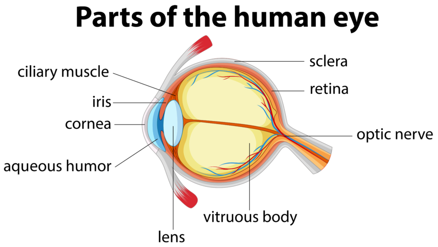
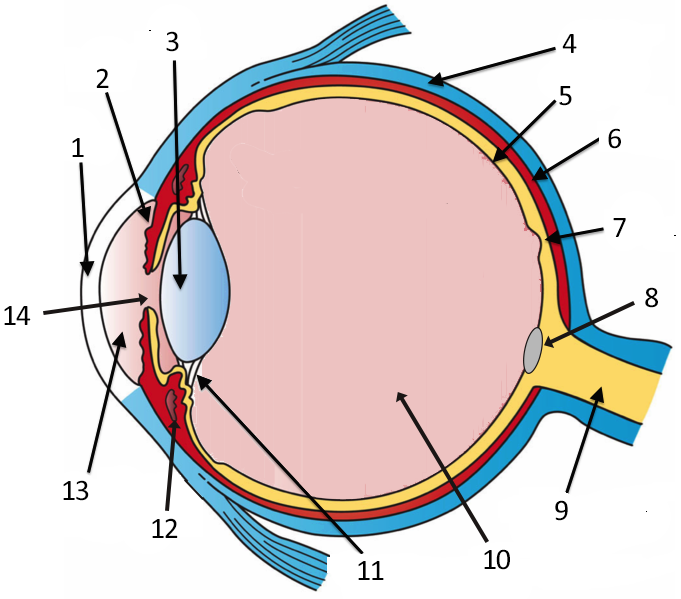
Parts of the human eye - labelling Google Classroom The diagram below points to different parts of the human eye. The human eye. Choose the correct labels for the parts shown. Choose all answers that apply: A is the crystalline lens. A A is the crystalline lens. B is the aqueous humour. B B is the aqueous humour. C is the iris. C C is the iris.

OUR EYES WORK LIKE CAMERA’S! Discovery Eye Foundation
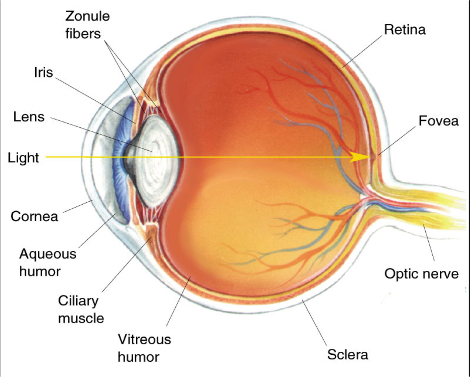
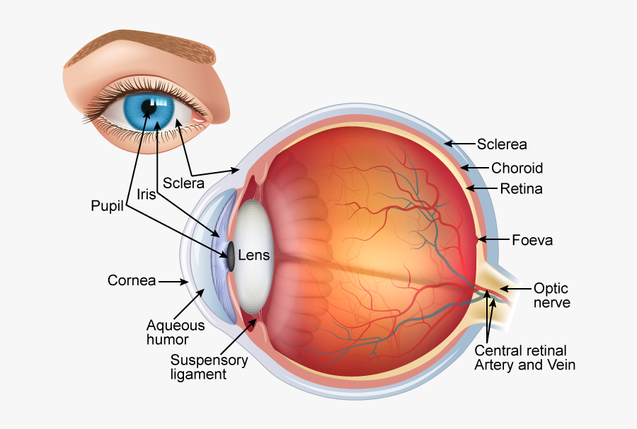
Light is focused primarily by the cornea - the clear front surface of the eye, which acts like a camera lens. The iris (colored part) of the eye functions like the diaphragm of a camera, controlling the amount of light reaching the retina by automatically adjusting the size of the pupil (aperture). The eye's crystalline lens is located.
HUMAN EYE (STRUCTURE, IMAGE FORMATION AND DIFFERENCE BETWEEN RODS AND CONES) « SimpleBiology
Label the parts of the human eye as quickly as possible! Quiz created specifically to line up with Miller/Levine Biology textbook, ISBN #0131152912. 9th and 10th grade Texas Biology. Enjoy!
Human Eye Different Parts and their functions Class 10 Teachoo
1. Conjunctiva The conjunctiva is the membrane covering the sclera (white portion of your eye). The conjunctiva also covers the interior of your eyelids. Conjunctivitis, often known as pink eye, occurs when this thin membrane becomes inflamed or swollen. Other eye disorders that affect the conjunctiva include:
Labeled Simple Labeled Human Eye Diagram
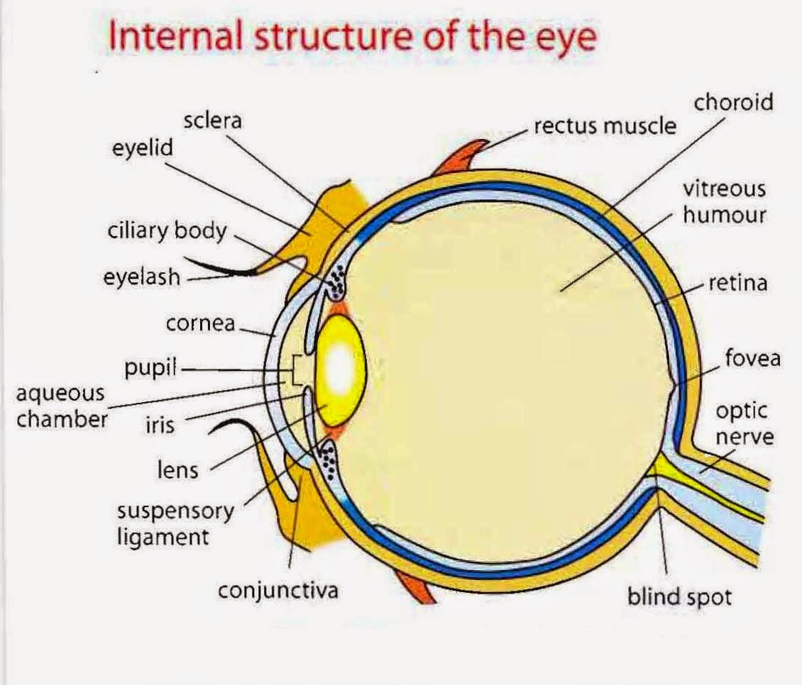
How do I use this Parts of the Eye Diagram Labelling Worksheet? In this resource, you'll find a 2-page PDF that is easy to download, print out, and use immediately with your class. The first page is a labelling exercise with two diagrams of the human eye. One is a view from the outside, and the other is a more detailed cross-section.
Eye Diagram Cliparts.co
Apr. 29, 2023 To understand the diseases and conditions that can affect the eye, it helps to understand basic eye anatomy. Here is a tour of the eye starting from the outside, going in through the front and working to the back. Eye Anatomy: Parts of the Eye Outside the Eyeball The eye sits in a protective bony socket called the orbit.
/GettyImages-695204442-b9320f82932c49bcac765167b95f4af6.jpg)
Structure and Function of the Human Eye
Chemistry Games. Periodic Table of the Elements, with Symbols. Periodic Table of the Elements. Periodic Table of the Elements, Period 1-3. Periodic Table of the Elements, Period 1-4. Periodic Table of the Elements, Period 4-5. Periodic Table of the Elements, Period 6-7. Periodic Table of the Elements, Other Nonmetals.
Vision and Eye Diagram How We See
Use this interactive labelling game to support children's learning about the parts of the human eye. Pupils drag and drop labels to identify the parts of the eye. A fun way to encourage independent reading and develop vocabulary and spelling. Share the resource via the unique PIN code generated by the Go! lesson builder so that they can all access the activity online on mobile devices.For more.
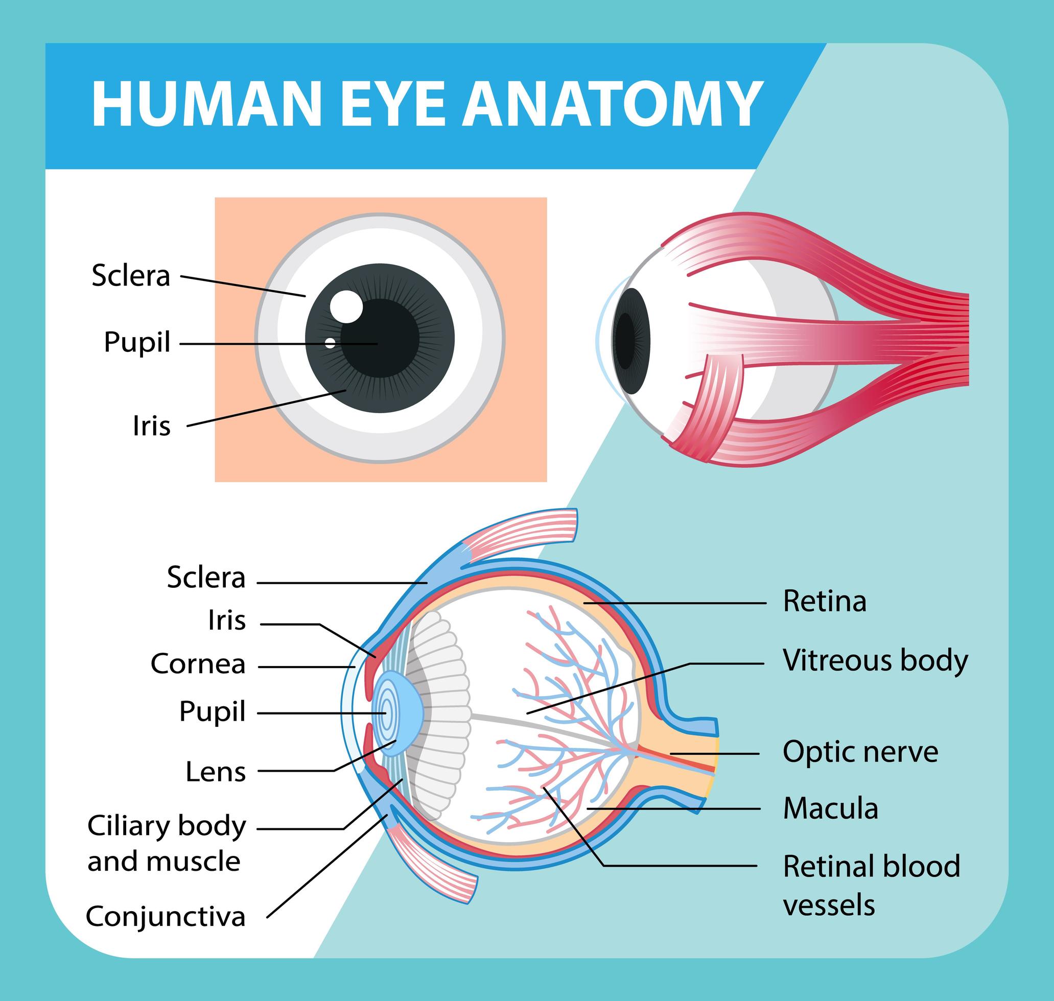
Diagram of human eye anatomy with label 1848847 Vector Art at Vecteezy
Human Eye Anatomy Can you locate the parts of the human eye? By smac17. 4m. 16 Questions. 337.1K Plays 337,142 Plays 337,142 Plays. Comments. Comments. Give Quiz Kudos. Give Quiz Kudos-- Ratings. hide this ad. Forced Order Answers have to be entered in order Answers have to be entered in order PLAY QUIZ Score.

Human Eye Anatomy, Structure and Function
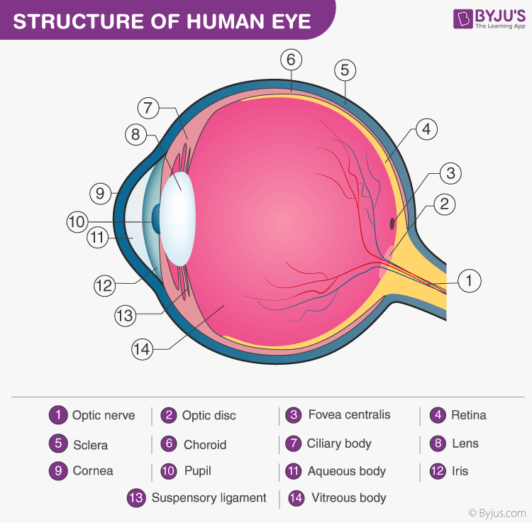
The inner layer of the eye is formed by the retina, the eye's light detecting component and the intraoptic part of optic nerve (cranial nerve II). The retina is made up of two separate functional units: the optic and nonvisual parts. The optic part of the retina is light-sensitive and consists of two layers: neural and pigmented layers.
Label the Eye
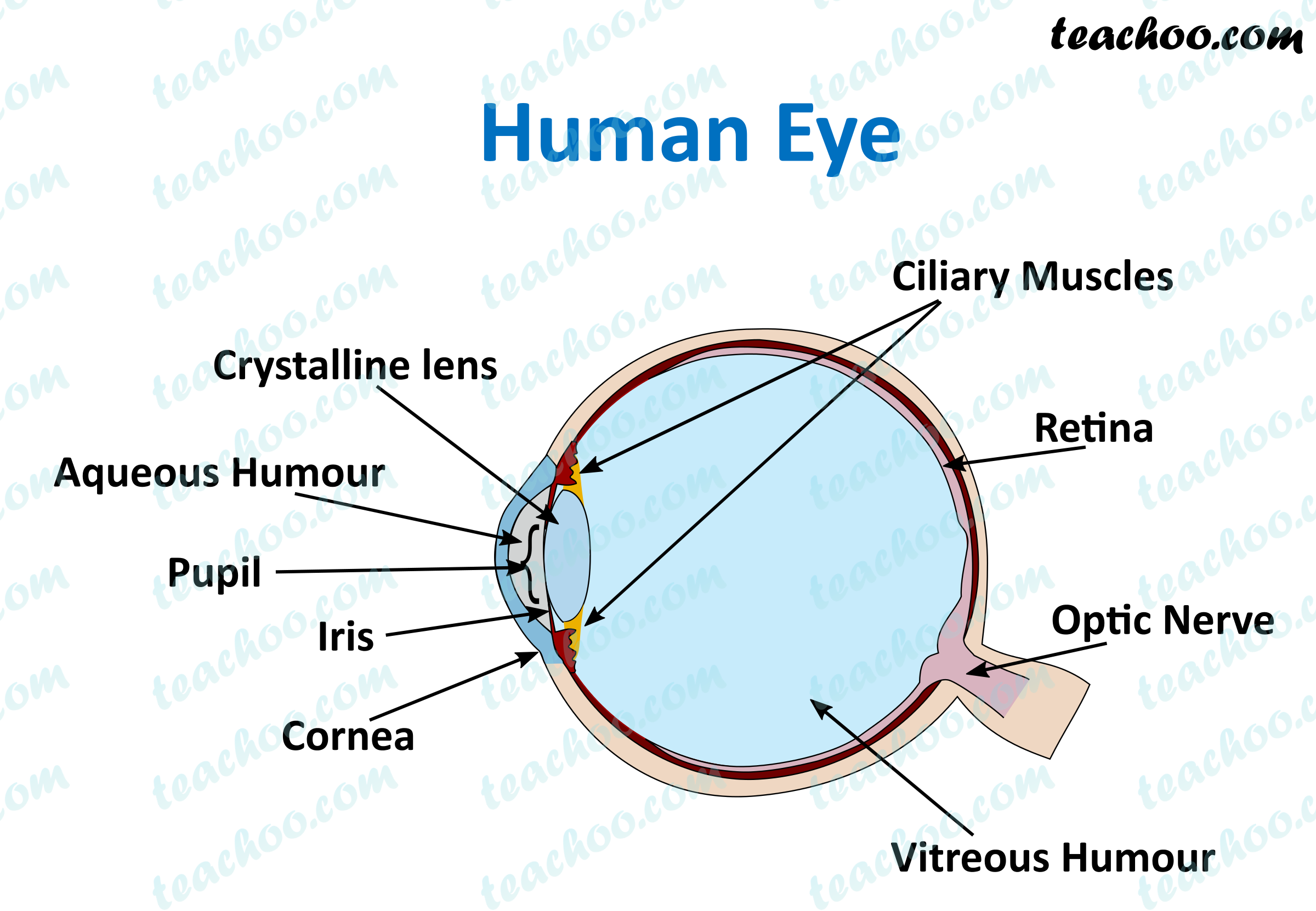
Anatomy of the Human Eye. Eyes are one of the most important organs of the body. A healthy pair of eyes means a clear vision, which plays a major role in day-to-day life and quality of experiences.
:max_bytes(150000):strip_icc()/eye-conjunctiva-871453538-5a26c6ad22fa3a0037d5edad.jpg)
How the Human Eye Works (Structure and Function)
Cornea: It is the transparent, anterior or front part of our eye, which covers the pupil and the iris. The main function is to refract the light along with the lens. Iris: It is the pigmented, coloured portion of the eye, visible externally. The main function of the iris is to control the diameter of the pupil according to the light source.

Eyelid Anatomy Diagram ANATOMY STRUCTURE
Aqueous humor - the clear, watery fluid inside the eye. It provides nutrients to the eye. Astigmatism - a condition in which the lens is warped, causing images not to focus properly on the retina. Binocular vision - the coordinated use of two eyes which gives the ability to see the world in three dimensions - 3D. Cones - cells the in the retina that sense color.
Human Eye Labelled Diagram , Free Transparent Clipart ClipartKey
Label the Eye 4.5 (29 reviews) Ciliary Body Click the card to flip 👆 Click the card to flip 👆 1 / 11 Flashcards Learn Test Match Created by beingdanny Teacher Students also viewed Q3: Label the Ear Teacher 9 terms gail-stowers Preview Labeling the Eye Teacher 16 terms MrsMeyer132 Preview Lymphatic System Med Terms Teacher 22 terms quizlette2671090

Human eye Extraocular Muscles Britannica
Label the Parts of the Eye by mrbahia 120,586 plays 11 questions ~30 sec English 11p 57 4.14 (you: not rated) Tries Unlimited [?] Last Played December 13, 2023 - 05:13 PM There is a printable worksheet available for download here so you can take the quiz with pen and paper. Remaining 0 Correct 0 Wrong 0 Press play! 0% 0:00.0 Other Games of Interest
