Anatomy of human foot with labels on white background — ankle, leg Stock Photo 200635434
Notes on Anatomy and Physiology Using Imagery to Relax the Weight
The midfoot is a pyramid-like collection of bones that form the arches of the feet. These include the three cuneiform bones, the cuboid bone, and the navicular bone. The hind foot forms the heel and ankle. The talus bone supports the leg bones (tibia and fibula), forming the ankle. The calcaneus (heel bone) is the largest bone in the foot.

Parts of the feet and legs Grammar Tips
The foot is the region of the body distal to the leg that is involved in weight bearing and locomotion. It consists of 28 bones, which can be divided functionally into three groups, referred to as the tarsus, metatarsus and phalanges. The foot is not only complicated in terms of the number and structure of bones, but also in terms of its joints.
Anatomy of human foot with labels on white background — ankle, leg Stock Photo 200635434
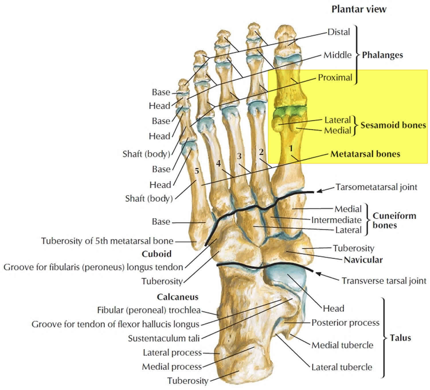
Foot bones and anatomy The human foot consists of 26 bones. These bones fall into three groups: the tarsal bones, metatarsal bones, and phalanges. Image credit: Stephen Kelly, 2019 Tarsal bones.
Advantage Orthopedic and Sports Medicine Clinic Gresham, OR Health Library
Joints Big toe Ball of foot Arch Heel General pain and swelling Speaking with a doctor Summary The location of pain in the foot can sometimes indicate the underlying cause. The cause will.

Foot Description, Drawings, Bones, & Facts Britannica
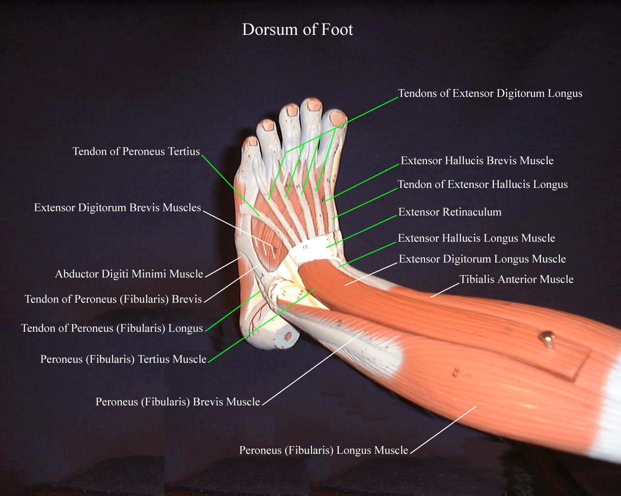
Cuboid Navicular Many of the muscles that affect larger foot movements are located in the lower leg. However, the foot itself is a web of muscles that can perform specific articulations that.
The 19 Muscles Of The Foot / The muscles of the foot Stock Image F001/4573 / The
The foot structure is complex, consisting of many bones, joints, ligaments and muscles. The foot is divided into three parts: rearfoot, midfoot, and forefoot. A clinician's ability to understand the anatomical structures of the foot is crucial for assessment and treatment, especially for clinicians working with clients with musculoskeletal conditions.
Turf toe causes, signs, symptoms, recovery, diagnosis & turf toe treatment
The foot is divided into three sections - the forefoot, the midfoot and the hindfoot. The forefoot This consists of five long bones (metatarsal bones) and five shorter bones that form the base of the toes (phalanges). The knuckles of the toes are called the metatarsophalangeal joint. The midfoot

Foot Anatomy 101 A Quick Lesson From a New Hampshire Podiatrist Nagy Footcare
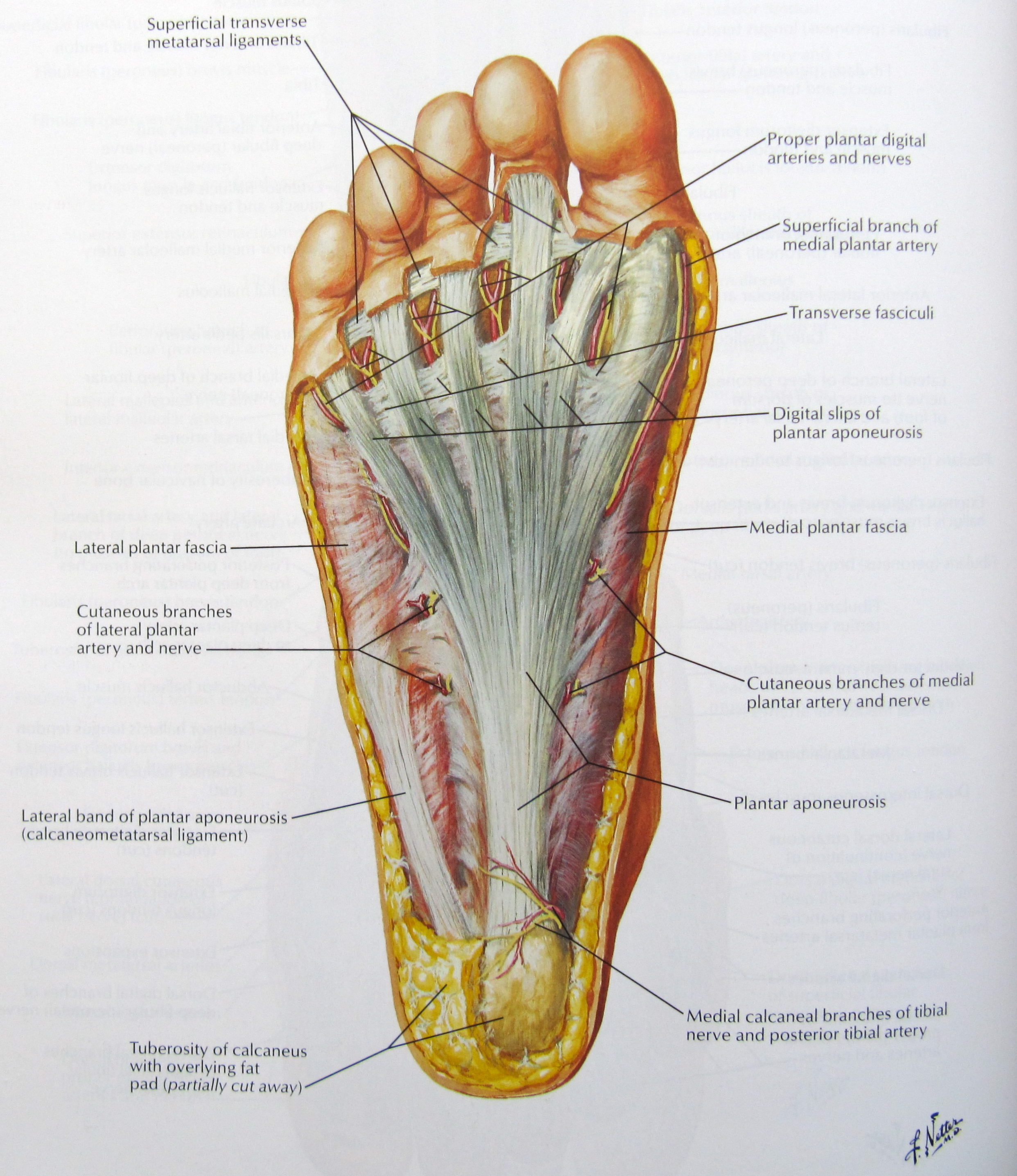
LABELED DIAGRAMS. Figure 1. Sections and Bones of the Foot A. Lateral (Left) B. Anterior (Right) Figure 2. Compartments of the Foot A. Cut Section through Mid-Foot. Figure 3. First Layer of the Foot A. Plantar View of Right Foot. Figure 4. Second Layer of the Foot A. Plantar View of Right Foot.

This chart shows foot and ankle bone and ligament anatomy, normal movement of the joints, and
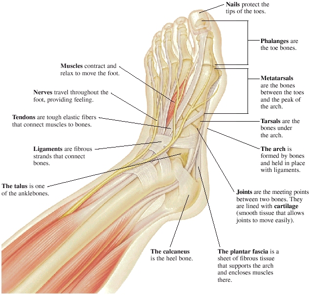
Foot Anatomy . There are many parts of the foot and all have important jobs. Each foot has 26 bones, over 30 joints, and more than 100 muscles, ligaments, and tendons. These structures work together to carry out two main functions:

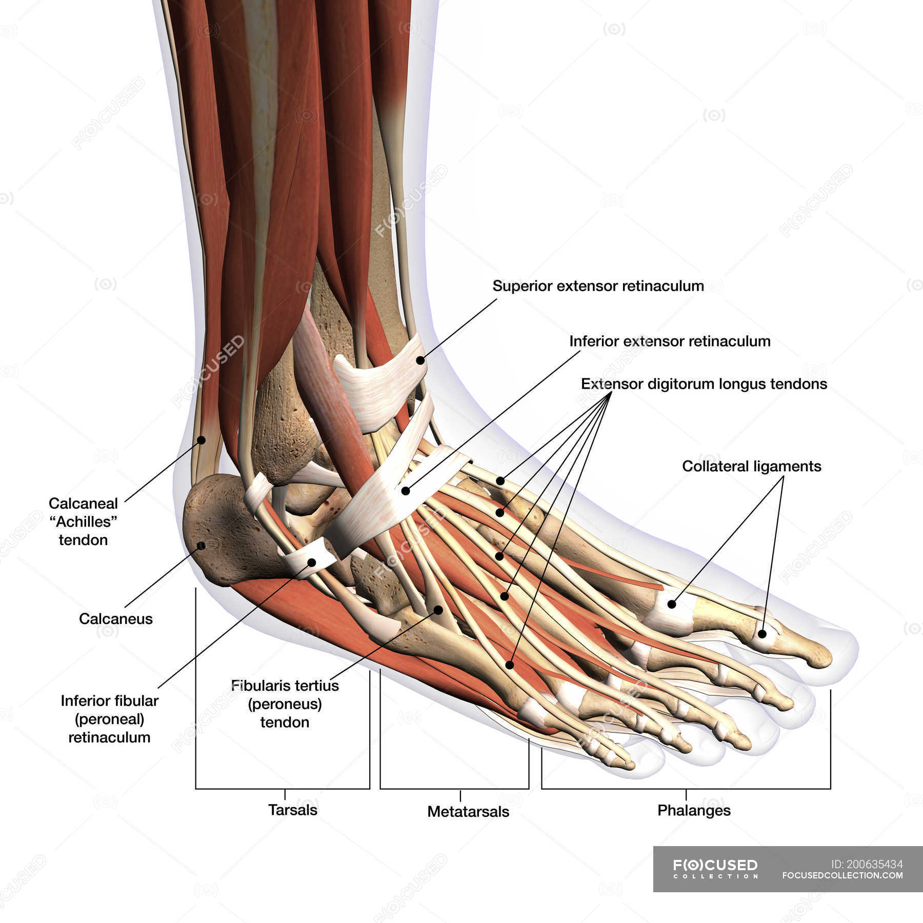
Foot & Ankle Bones
Did you know that the human foot has 26 bones, 33 joints, and over 100 muscles, tendons, and ligaments? It's a complex structure that plays a vital role in our everyday lives. In this blog post, we will explore the different parts of the foot and what they do. We'll also discuss common injuries and conditions that can affect the foot.

Chart of FOOT Dorsal view with parts name Vector image Stock Vector Image & Art Alamy
In most two-footed and many four-footed animals, the foot consists of all structures below the ankle joint: heel, arch, digits, and contained bones such as tarsals, metatarsals, and phalanges; in mammals that walk on their toes and in hoofed mammals, it includes the terminal parts of one or more digits. a dog's feet
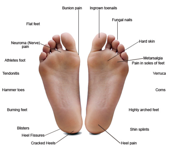
Common Foot Problems — Hawaii Podiatry
There are a variety of anatomical structures that make up the anatomy of the foot and ankle (Figure 1) including bones, joints, ligaments, muscles, tendons, and nerves. These will be reviewed in the sections of this chapter. Figure 1: Bones of the Foot and Ankle Regions of the Foot

Image result for skull sketch anatomy underside Foot anatomy, Human anatomy picture, Anatomy
The foot is one of the most complex parts of the body. It consists of 28 bones connected by many joints, muscles, tendons, and ligaments. The foot is prone to many types of injuries. Foot pain and problems can cause pain and inflammation, limiting movement. Muscles contract and relax to move the foot.
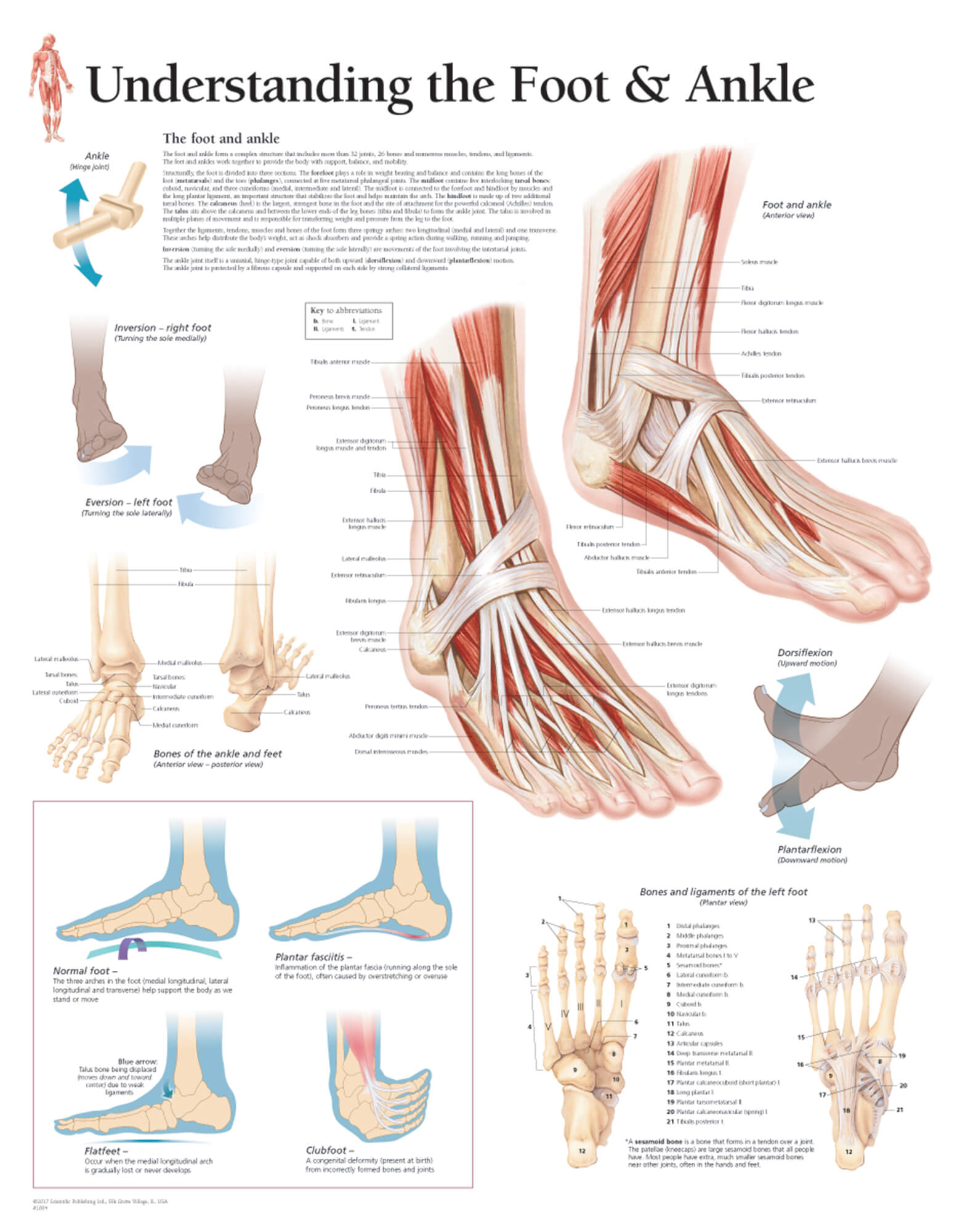
Understanding the Foot & Ankle Scientific Publishing
Ligaments are fibrous strands that connect bones. Nerves travel throughout the foot, providing feeling. Nails protect the tips of the toes. Phalanges are the toe bones. Metatarsals are the bones between the toes and the ankle bones. Tarsals are bones of the rear foot (hindfoot) or middle foot (midfoot). The talus is one of the ankle bones.

Foot pain looking up the chain
33 joints more than 100 muscles, tendons, and ligaments Bones of the foot The bones in the foot make up nearly 25% of the total bones in the body, and they help the foot withstand weight..
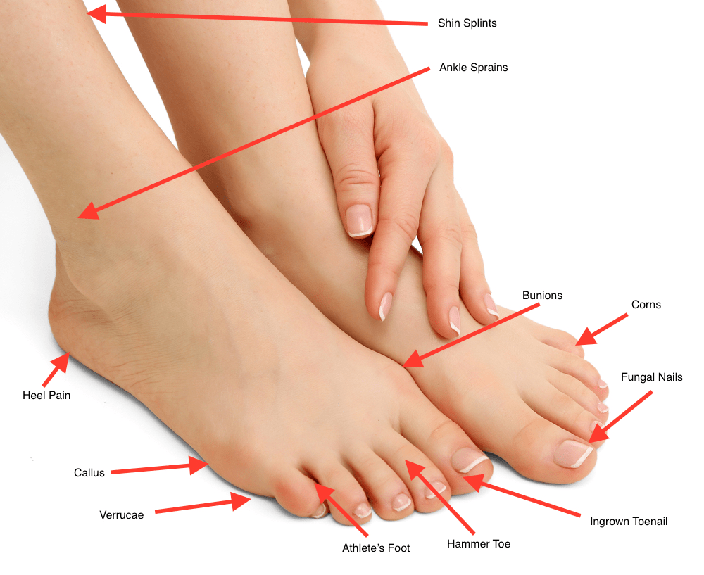
Common Foot Problems Cornwall's Leading Foot Clinic Chiropodist
There are 26 bones in the foot, divided into three groups: Seven tarsal bones Five metatarsal bones Fourteen phalanges Tarsals make up a strong weight bearing platform. They are homologous to the carpals in the wrist and are divided into three groups: proximal, intermediate, and distal.
