UCSD Musculoskeletal Radiology

Добавочные кости Портал радиологов
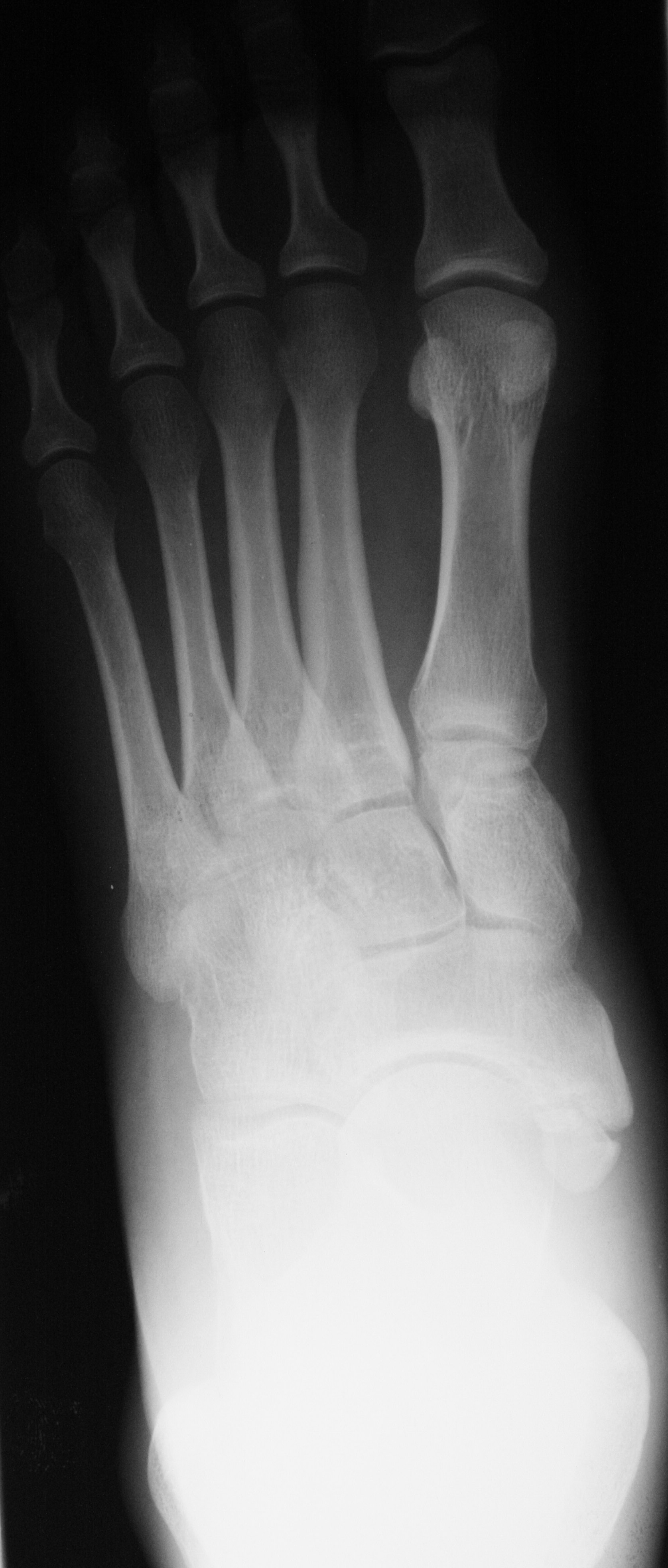
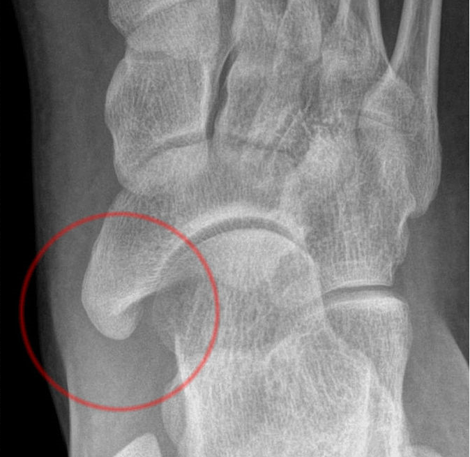
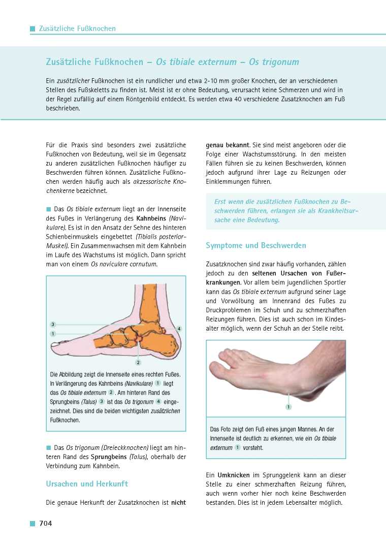
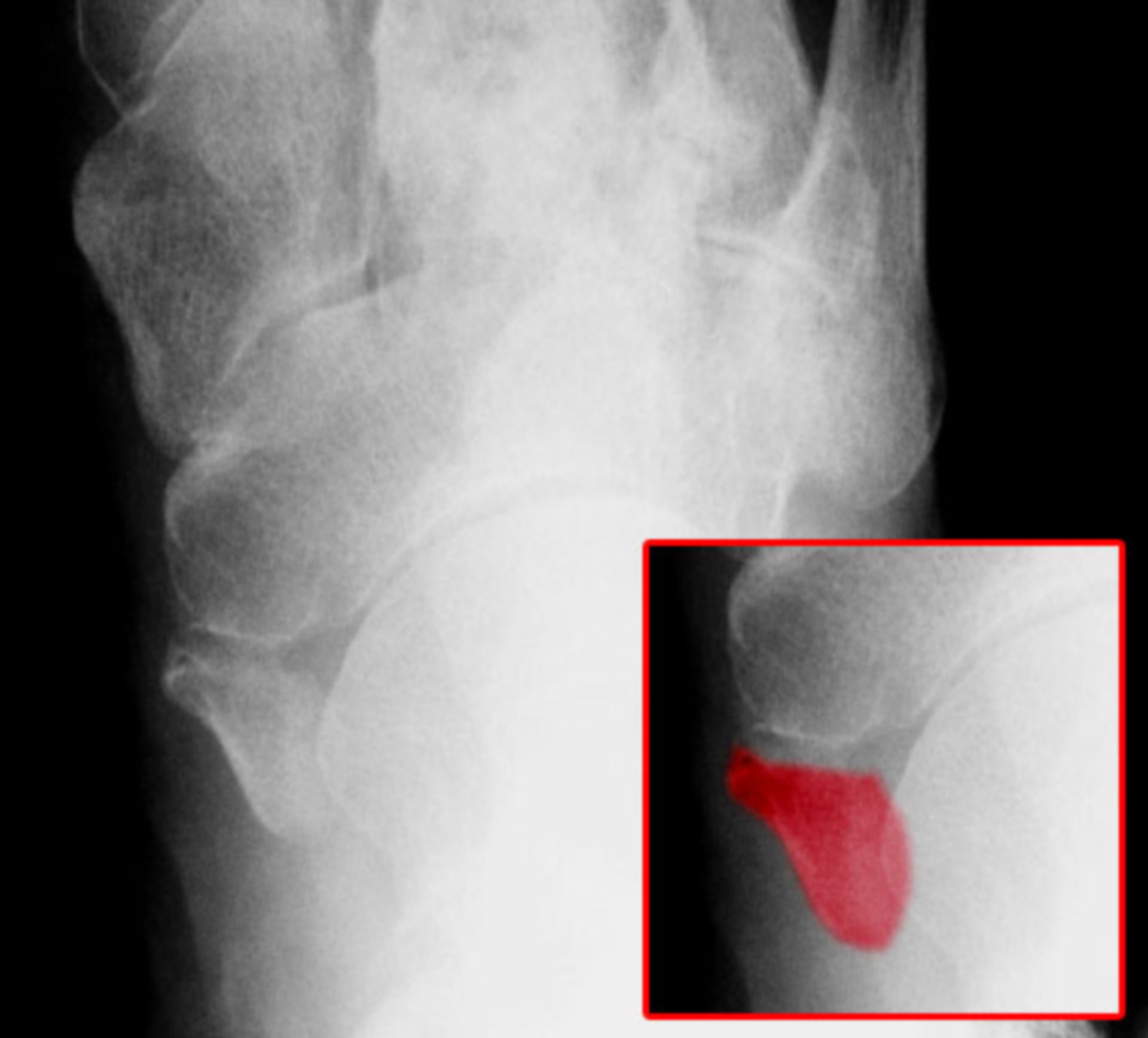
The os tibiale externum(1) also called the accesory navicular is the most commonly found accesory ossicle of the foot with reported incidence of about 25-30%. It is located on the posteromedial aspect of the foot adjacent to the posteromedial tuberosity of the navicular bone. Three types of accessory tibiale externum have been described in.

Os_tibiale_externum Don't the Bubbles
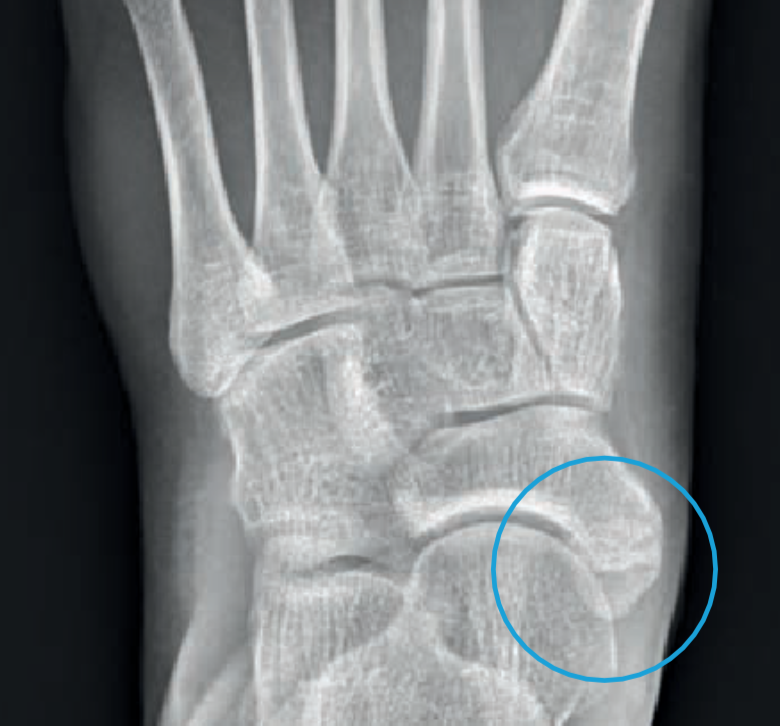
Classification. The Geist classification divides the accessory navicular bones into three types. Type 1: An os tibiale externum is a 2-3 mm sesamoid bone in the distal posterior tibialis tendon.Usually asymptomatic. Type 2: Triangular or heart-shaped ossicle measuring up to 12 mm, which represents a secondary ossification center connected to the navicular tuberosity by a 1-2 mm layer of.
UCSD Musculoskeletal Radiology
Also known as 'os tibiale externum' or 'os navicularum', accessory navicular syndrome refers to a congenital abnormality related to the growth of an extra bone within the foot. This additional piece of bone is not present in a normal human foot and grows toward the middle inner part of the foot near the navicular bone.

Os tibiale externum sagittal T2 YouTube
Also known as os naviculare or os tibiale externum, an accessory navicular is an extra bone on the inside of the navicular (the bone in the middle of the arch of the foot) and within the posterior tibial tendon that attaches to the navicular bone. Top-view of accessory navicular in the right foot.
Os Tibiale Externum Ortobas
Citation, DOI, disclosures and article data. Accessory ossicles of the feet are common developmental variants with almost 40 having been described. Some of the more common include 1-4: os peroneum. os subfibulare. os subtibiale. os tibiale externum (accessory navicular) os trigonum. os calcaneus secundaris.
Orthopädie für Patienten Zusätzliche Fußknochen Os tibiale externum
The os intermetatarseum is less common than the os tibiale externum, os trigonum, and os peroneum. The estimated prevalence is 1.2%-10% [ 2 , 9 ]. Reichmister, et al. reported three cases of painful os intermetatarseum, and described compression of the deep peroneal nerve by the os intermetatarseum [ 23 ].

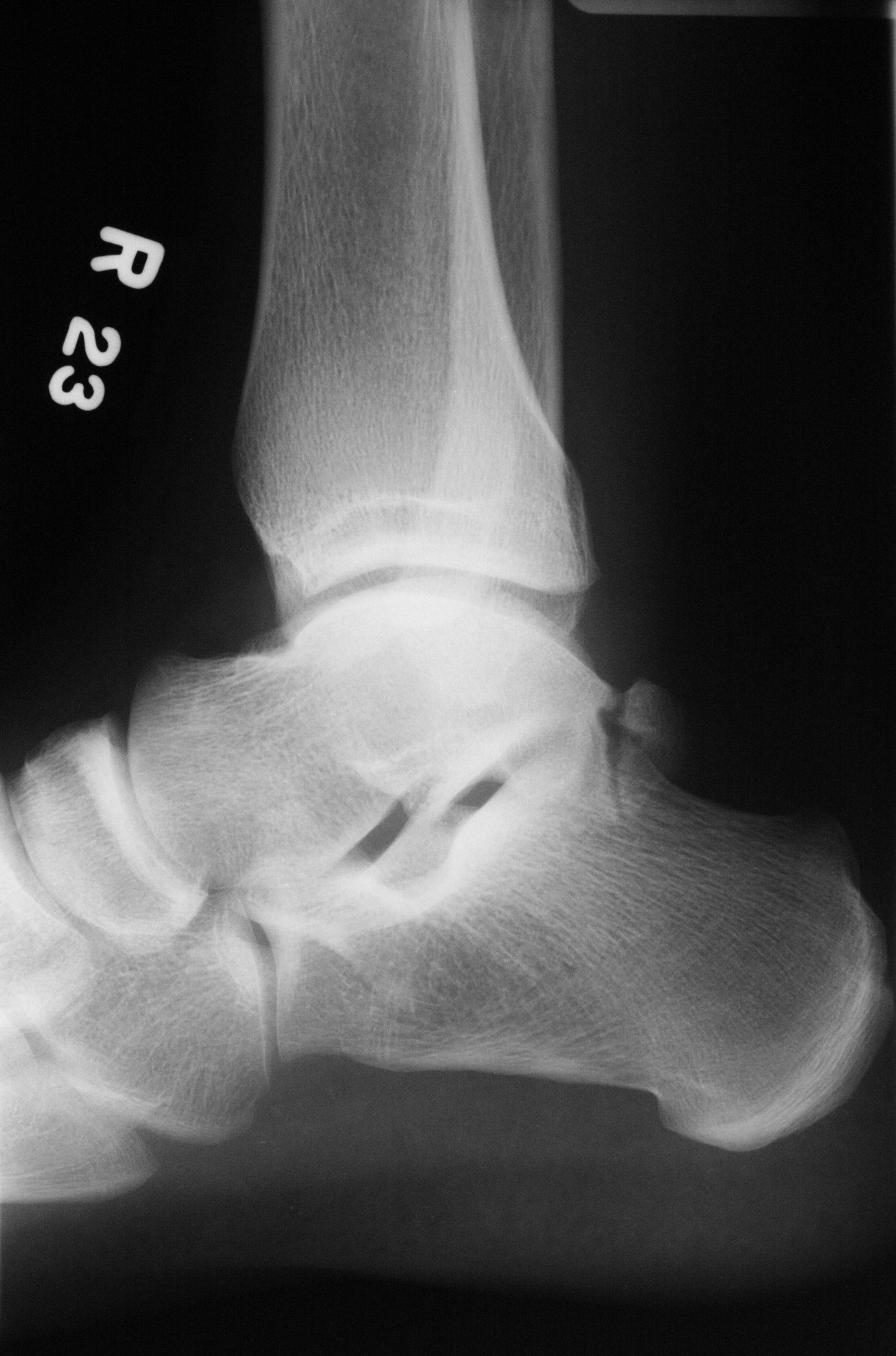
Os tibiale externum type II Image
The Geist1 classification divides accessory navicular bones into three types: type 1 accessory navicular bone. also known as os tibiale externum. 2-3 mm sesamoid bone embedded within the distal portion of the posterior tibial tendon. no cartilaginous connection to the naviculam tuberosity and may be separated from it by up to 5 mm.

Schmerzhaftes Os tibiale externum Dr.medic Manuel Nastai
The accessory navicular syndrome, also known as os naviculare syndrome occurs when a type II accessory navicular becomes painful due to movement across the pseudo-joint between the ossicle and the navicular bone.. Radiographic features Ultrasound. It can be inferred on musculoskeletal ultrasound if a patient's pain is located at a type II accessory navicular and the patient is tender to.

Treating The Accessory Navicular In Young Athletes
The accessory navicular (os navicularum or os tibiale externum) is an extra bone or piece of cartilage located on the inner side of the foot just above the arch. It is incorporated within the posterior tibial tendon, which attaches in this area. An accessory navicular is congenital (present at birth).

Os tibiale externum Geist classification Radiology Case
The os tibiale externum is also known as accessory navicular bone, os naviculare secundarium, accessory (tarsal) scaphoid, or prehallux. It is found within the tibialis posterior tendon near its insertion on the navicular bone. The os peroneum is a small sesamoid bone located within the peroneus longus tendon, adjacent to the cuboid.
Os tibiale externum DocCheck
The accessory navicular (os navicularum or os tibiale externum) is an extra bone or piece of cartilage located on the inner side of the foot just above the arch. It is incorporated within the posterior tibial tendon, which attaches in this area and can lead to Accessory Navicular Syndrome. An accessory navicular is congenital (present at birth.

Surgery Assistant
Os tibiale externum; Pirie's bone; Talonaviculare ossicle; Os scaphoideum accessorium; URL of Article. An accessory navicular is a large accessory ossicle that can be present adjacent to the medial side of the navicular bone. The tibialis posterior tendon often inserts with a broad attachment into the ossicle. Most cases are asymptomatic but.
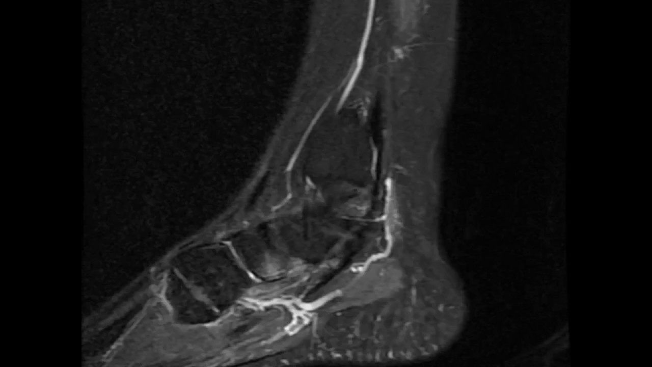
Os tibiale externum sagittal T2 fat sat YouTube
accessory navicular (os tibiale externum) os intermetatarseum. Most common sesamoids. os peroneum. located in the peroneus longus tendon. hallux sesamoids. located in the flexor hallucis brevis tendon at the base of the 1st metatarsal head. Classification. Accessory Ossicles and Sesamoids of the Foot and Ankle.

Roentgen Ray Reader Types of Accessory Navicular Bones
Os tibiale externum (accessory navicular) is a large ossicle adjacent to the medial side of the navicular bone. The tibialis posterior tendon often inserts with a broad attachment onto the ossicle, which may cause a painful tendinosis due traction between the ossicle and the navicular. Such changes are best seen on MRI.
Knickfuss Leonardo
Accessory Navicular. Acessory Navicular is a common idiopathic condition of the foot that presents with an enlargement of the navicular bone. Diagnosis is made with plain radiographs of the foot showing a plantar medial enlargement of the navicular bone. Treatment is generally conservative with shoe modifications and a short period of cast.
UCSD Musculoskeletal Radiology
Os tibiale externum (OTE) also termed accessory navicular, os naviculare, or os navicularis is a common accessory bone in the foot located medial and sometimes proximal to the navicular tuberosity. It is attached and continuous with the tibialis posterior tendon and is present in 10 to 15% of the population either unilateral or bilateral.
