Pin on Equine Anatomy
horse anatomy on Pinterest Animal Anatomy, Horses and Anatomy
Dr Christin Finn, DVM, CVA details the underlying skeleton and muscle structure of a horse by using a live painted horse Find more information about your hor.
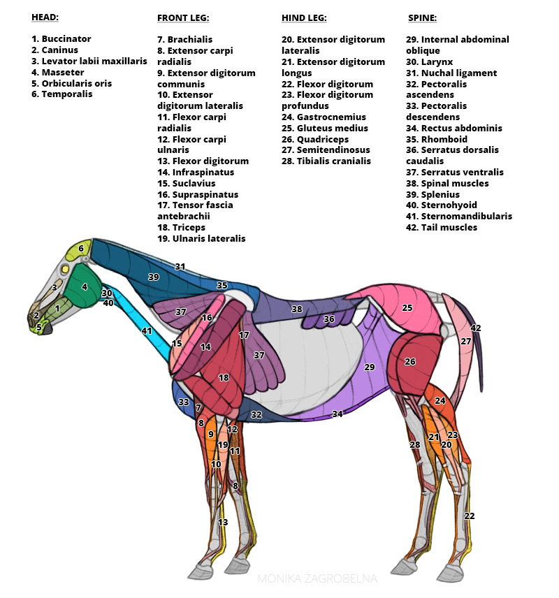
Horse Anatomy for Artists Skeleton and Muscle Diagrams Monika Zagrobelna
Tibia. Large and only weight bearing component of crus (stifle/ knee) Large tibial tuberosity - patellar ligament. Medial tibia is subcutaneous. Cochlea is inclined craniolaterally. This causes the lower limb to move laterally on flexion. Fibula is greatly reduced. Distally incorporated into tibia. Proximally tightly articulated with tibia.

Pin on Caballos
Tendons are tough bands of connective tissue made up mostly of a protein called collagen. They stretch very little. Tendons attach each end of a muscle to a bone. At selected points, they are located within sheaths that allow them to move easily. Ligaments are also tough cords formed of connective tissue.

Pin on Equine Anatomy
Head and Neck - Horse Muscle Anatomy. The muscles of the head and neck allow the horse to flex, extend and tilt its head. Key muscles include: Temporalis - raises the jaw. Masseter - closes the jaw. Sternomandibularis - opens the mouth by lowering the jaw. Brachiocephalicus - flexes the head and neck; turns the head side to side.
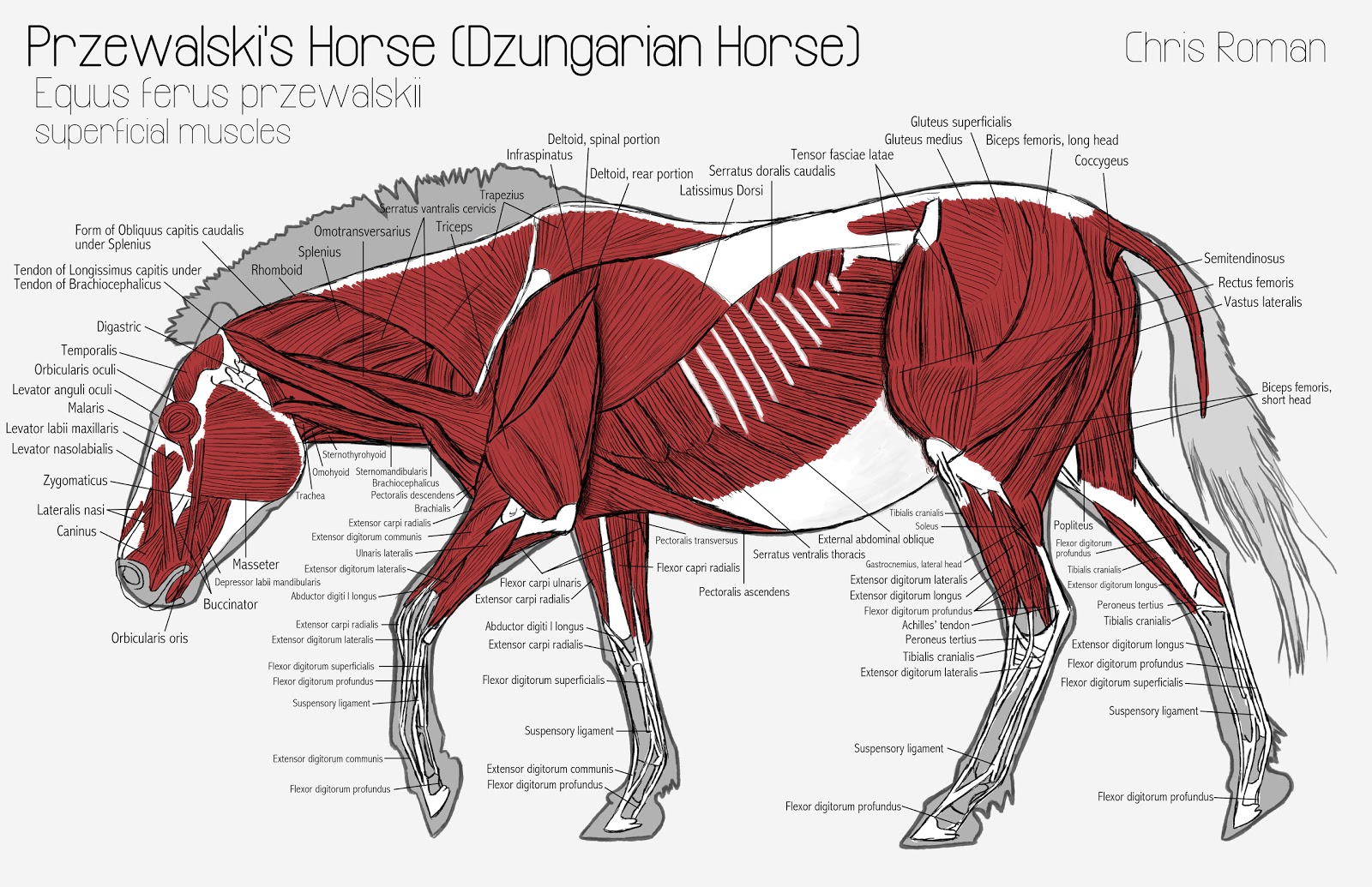
Chris Roman Horse Anatomy Study
Anatomy. The Iliopsoas muscle group is comprised of the Psoas Major and the Iliacus muscle. Psoas Major.. Rock, S. (2012) Anatomy of the Horse. Posted by Vet Physio Phyle November 12, 2018 October 30, 2020 Posted in Uncategorized Tags: muscle Post navigation. Previous Post Previous post: Persevering through physical illness. Next Post Next.

Anatomy of the horse The shoulder Animal Osteo Louise Mauferon
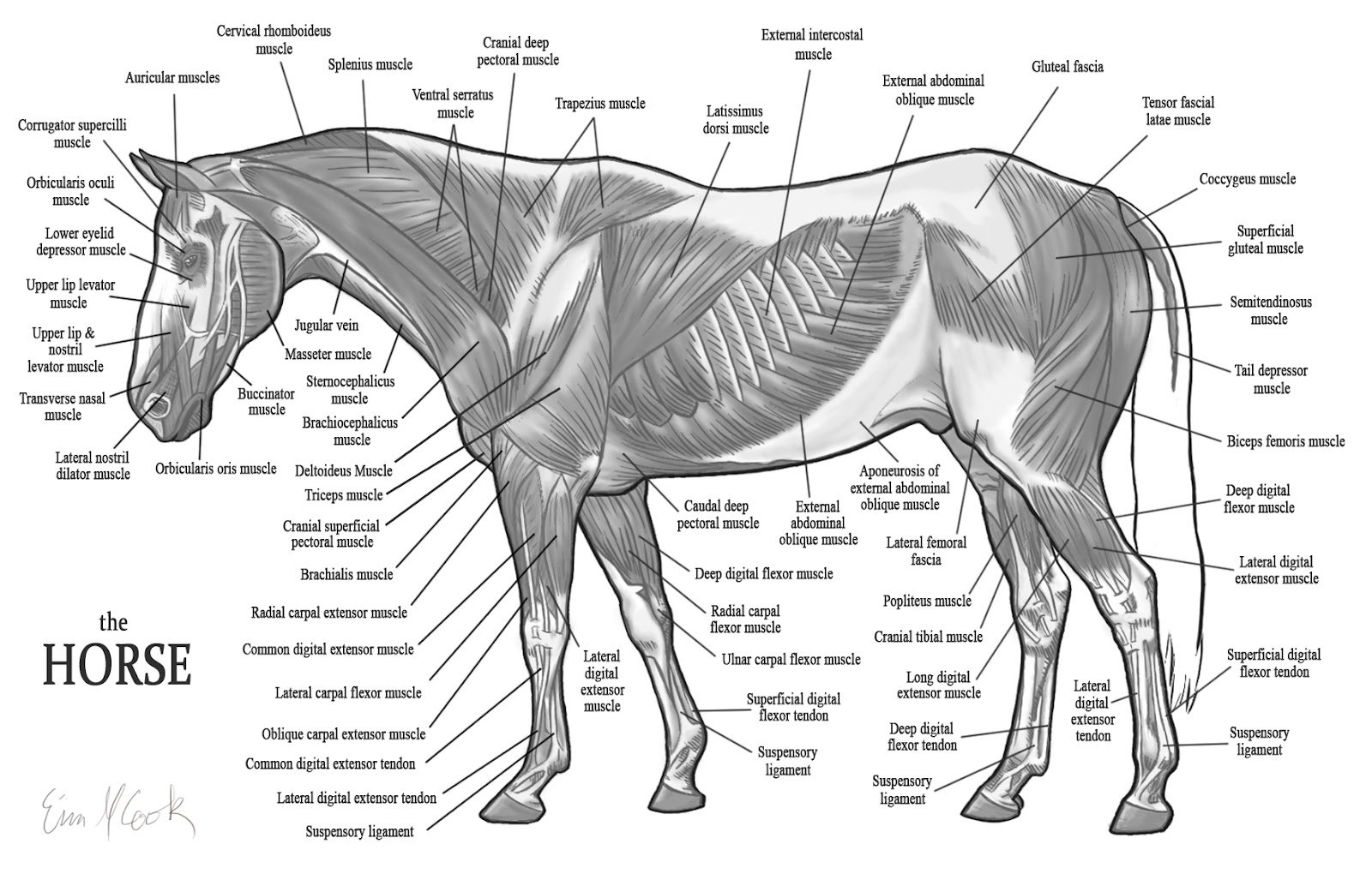
The horse's body possesses approximately 700 muscles that control movement. By understanding how horses' muscles contract to produce movement, you can formulate training and rehab strategies.

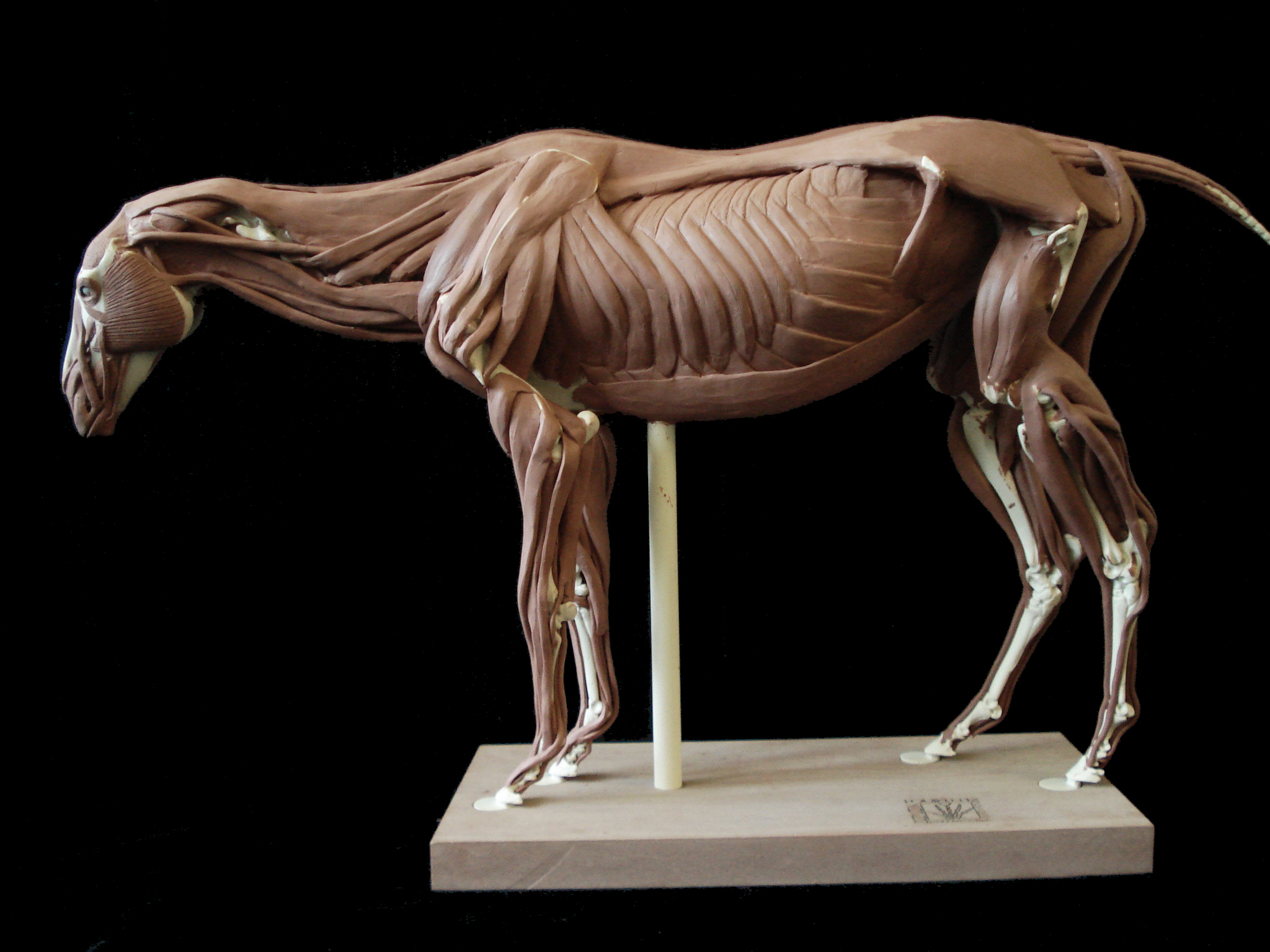
horse anatomy model Google Search Horse anatomy, Horse health, Horses
Points of a horse. Equine anatomy encompasses the gross and microscopic anatomy of horses,. the tongue and related muscles, the cheeks and the lips. Horses also have three pairs of salivary glands, the parotoid (largest salivary gland and located near the poll), mandibular (located in the jaw), and sublingual (located under the tongue).

Horse anatomy, Horse behavior, Horses
Horse muscle anatomy chart Horse anatomy skeleton Horse head. The horse's head is quite heavy - it can weigh approximately 16 kg if the horse is big. You should remember about this, especially when you allow the horse to "hang" on a bit and you "carry" his head's weight with your arms throughout the whole ride by holding the reins. After an.

Horse Muscular Anatomy Poster
Wing of atlas and mastoid process of temporal bone. Elevates head and neck. Bends head and neck laterally. Stabilizes and extends vertebral column. Dorsal branch of local spinal nerve. M. Semispinalis capitis. Articular processes of C2/3-7 and transverse processes of T1-6/7. Occipital bone. Elevates head and neck.
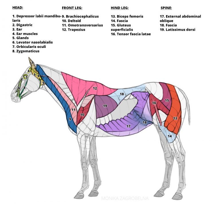
Horse Anatomy for Artists Skeleton and Muscle Diagrams Monika Zagrobelna
The horse's musculoskeletal system consists of the bones, cartilage, muscles, ligaments, and tendons. Their primary function is to support of the body, provide motion, and protect vital organs. There are 205 bones in the horse's skeleton. Twenty of these bones are in each foreleg and 20 in each hind limb, for a grand total of 80 bones in.
Horse muscle anatomy
Abscess - An infection of the sensitive tissues of the hoof. Cause: Rocks, deep bruising of the sole, nails, and/or sharp objects that puncture sensitive tissue of the hoof. Abscesses will follow the path of least resistance. Sometimes puncture hole may be seen (black spot). The leg may be hot and swollen. Call a vet if not experienced!

Full color poster of the equine deep muscular system. Perfect for a
Muscles of the forehand. Opens and closes the jaw. Allows chewing. movement. Too strong a. forward, raises it in collection, swings the foreleg forward. rein contact stops free forward movement. Moves the head and neck. Over developed in ewe or bull necked horses, difficult to get into a relaxed shape.

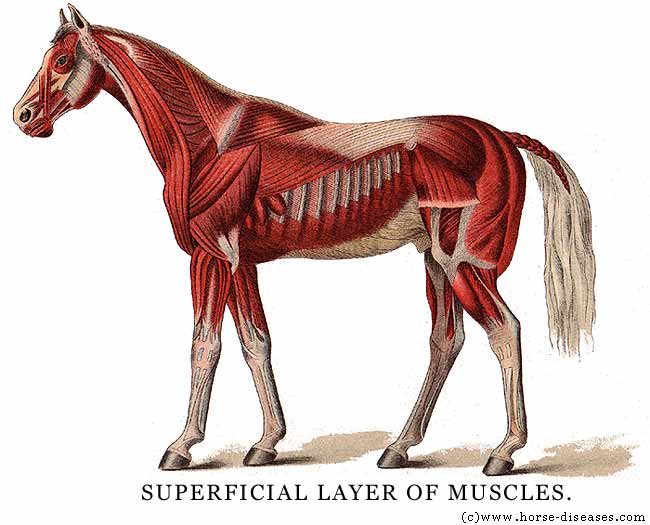
THE ACTION OF MUSCLES
The top line is a horse muscle anatomy term that describes the coverage of the horse trapezius muscle, neck, back withers, loin, and coup. Horses with a muscular topline will be stronger and more athletic than those without one. Protein is a natural horse muscle supplement and one of the best horse supplements for muscle gain.
The Equus Ally The Basics Horse Anatomy
Horse Anatomy and Muscle Diagrams. This page contains color coded pictures of the horse's deep, superficial and hind end muscles.. Like humans, all horses need to have their muscles stretched to prevent injury and improve their comfort and performance level. We created this 25 video collection to teach you how to safely and correctly do this!
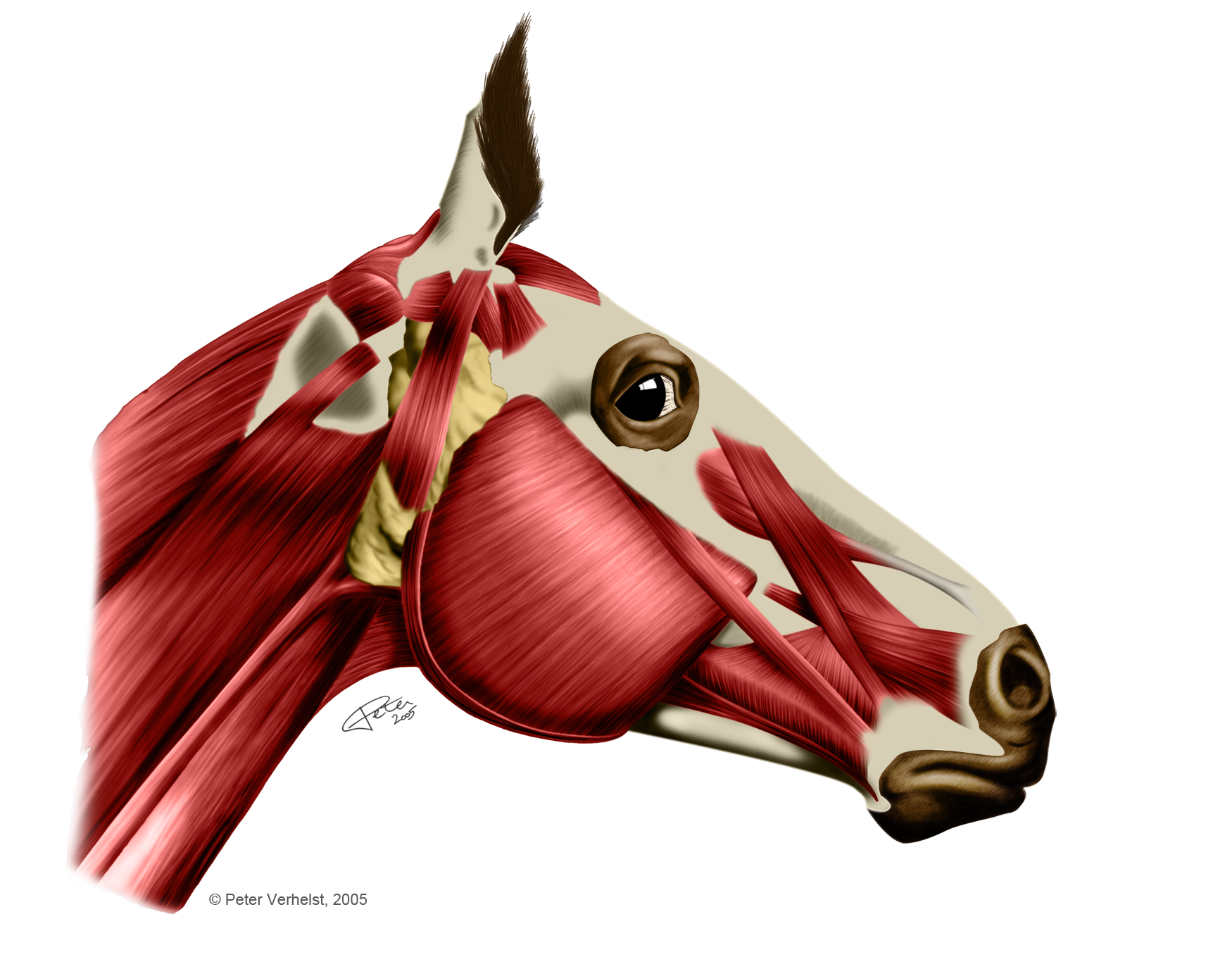
Muscles of a horse head by PeterAndTheWolf on DeviantArt
Types of muscle. There are 3 types of muscle, all found within the equine: Skeletal muscle: Contraction of these muscles leads to the muscle pulling a tendon, which in turn pulls a bone. Moving a bone results in either flexing or extending a joint.

Image result for horse muscles diagram Horse anatomy, Horses, Horse
The Gaskin is an important muscle on the inside of a horse's leg, just above the hock 1:. That's a lot of information for basic horse anatomy. However, this knowledge can help provide better care for your horse residents and give staff a good foundation to build from. This resource is far from extensive and a "Part 2" will be in the.
