Microscope diagram Tom Butler Technical Drawing and Illustration Projects Pinterest
Labelled Diagram of Microscope Parts
Create a poster that labels the parts of a microscope and includes descriptions of what each part does. Click "Start Assignment". Use a landscape poster layout (large or small). Search for a diagram of a microscope. Using arrows and textables label each part of the microscope and describe its function. More options.
Parts Parts And Functions Of A Microscope
Introduction If you meet some cell biologists and get them talking about what they enjoy most in their work, you may find it comes down to one thing: secretly, they're all microscope freaks.

Parts of a microscope with functions and labeled diagram
Fluorescence microscopes: These use fluorescent dyes to highlight specific structures or molecules in a sample and are commonly used in biological research. X-ray microscopes: These use X-rays to produce images of the internal structure of samples and are often used to study materials and biological specimens.

Parts of a Microscope The Comprehensive Guide Microscope and Laboratory Equipment Reviews
The working principle of a simple microscope is that when a lens is held close to the eye, a virtual, magnified and erect image of a specimen is formed at the least possible distance from which a human eye can discern objects clearly. Magnification formula The magnification power of a simple microscope is expressed as: M = 1 + D/F Where
Parts Of A Microscope Worksheet
Parts of the Microscope (Labeled Diagrams) By Editorial Board December 14, 2022 The microscope is one of the must-have laboratory tools because of its ability to observe minute objects, usually living organisms that cannot be seen by the naked eyes. It is categorized into two: simple and compound microscopes.
Cells and Microscopes
Use this interactive to identify and label the main parts of a microscope. Drag and drop the text labels onto the microscope diagram.

301 Moved Permanently
Light and electron microscopes allow us to see inside cells. Plant, animal and bacterial cells have smaller components each with a specific function. We need microscopes to study most cells.

Microscope Diagram Labeled, Unlabeled and Blank Parts of a Microscope
A Study of the Microscope and its Functions With a Labeled Diagram - Science Struck A Study of the Microscope and its Functions With a Labeled Diagram To better understand the structure and function of a microscope, we need to take a look at the labeled microscope diagrams of the compound and electron microscope.

5 Types of Microscopes with Definitions, Principle, Uses, Labeled Diagrams
Labeling the Parts of the Microscope This activity has been designed for use in homes and schools. Each microscope layout (both blank and the version with answers) are available as PDF downloads. You can view a more in-depth review of each part of the microscope here. Download the Label the Parts of the Microscope PDF printable version here.
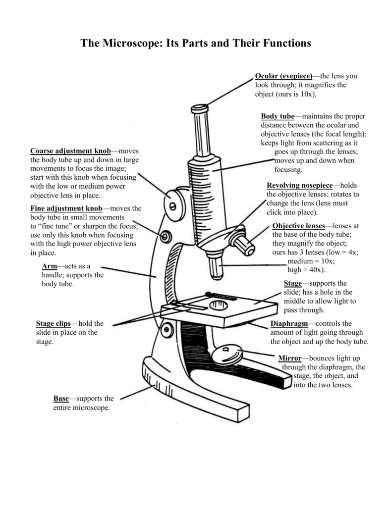
The Microscope Its Parts and Their Functions
A microscope is a piece of laboratory optical equipment used to magnify small things that are too small for the details to be seen by the naked eye. The microscope is the microbiologist's most basic tool, and every microbiology student needs some background knowledge on parts of a microscope and how microscopes work.
1.5 Microscopy Biology LibreTexts
A labeled diagram of microscope parts furnishes comprehensive information regarding their composition and spatial arrangement within the microscope, enabling researchers to comprehend their function effectively. In this comprehensive article, we will delve into the intricate parts of the microscope, exploring their functions in detail..
Clipart microscope parts labeled WikiClipArt
So, a compound microscope with a 10x eyepiece magnification looking through the 40x objective lens has a total magnification of 400x (10 x 40). Specimen or slide: The object used to hold the specimen in place along with slide covers for viewing.. Compound Microscope Parts, Functions, and Labeled Diagram. Parts of a Compound Microscope.
Ag Biology Unit 2
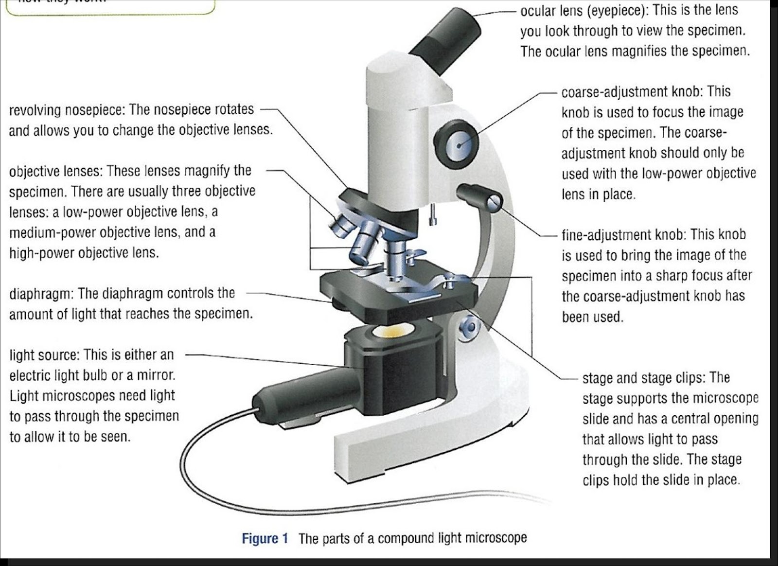
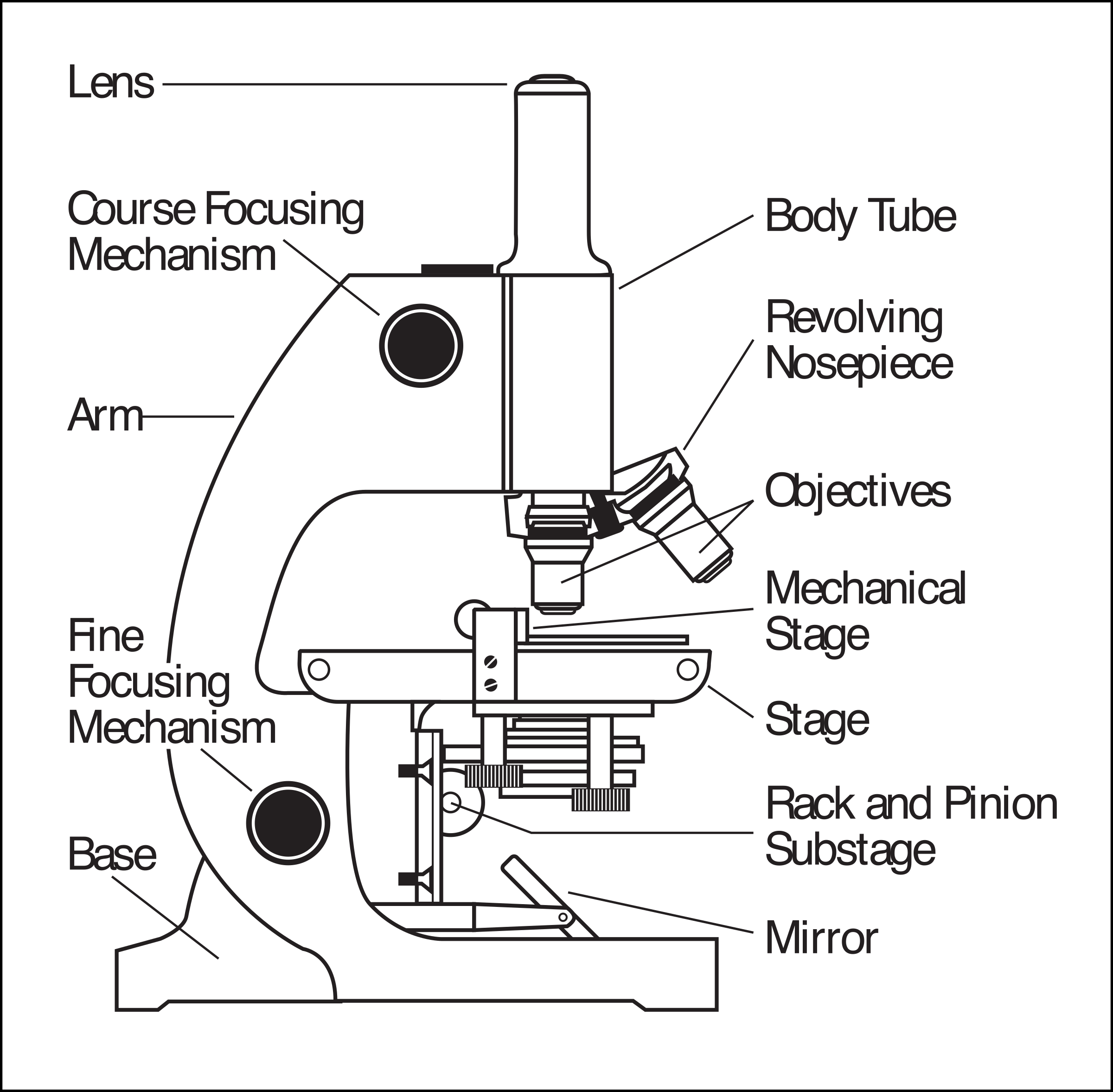
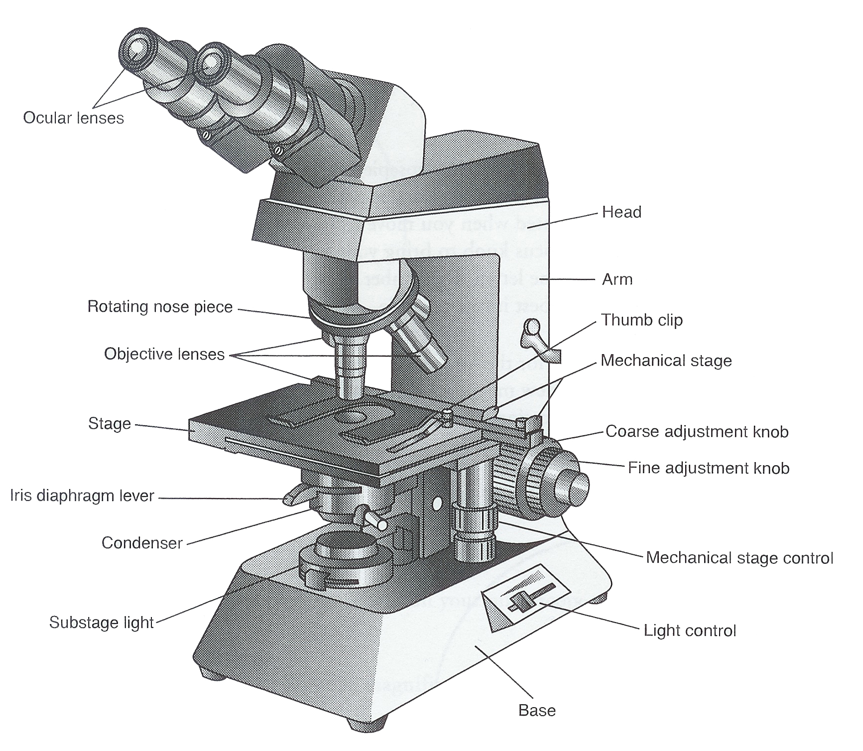
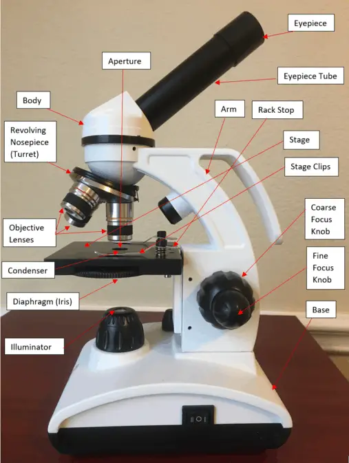
Figure: Diagram of parts of a microscope. There are three structural parts of the microscope i.e. head, arm, and base. Head - The head is a cylindrical metallic tube that holds the eyepiece lens at one end and connects to the nose piece at other end. It is also called a body tube or eyepiece tube.
16 Parts of a Compound Microscope Diagrams and Video Microscope Clarity
Labeled diagram of a compound microscope Major structural parts of a compound microscope Optical components of a compound microscope Eyepiece Eyepiece tube Objective lenses Nosepiece Specimen stage Coarse and fine focus knobs Rack stop Illuminator Condenser Abbe condenser Iris Diaphragm Condenser Focus Knob Summary An overview of microscopes
How to Use a Microscope
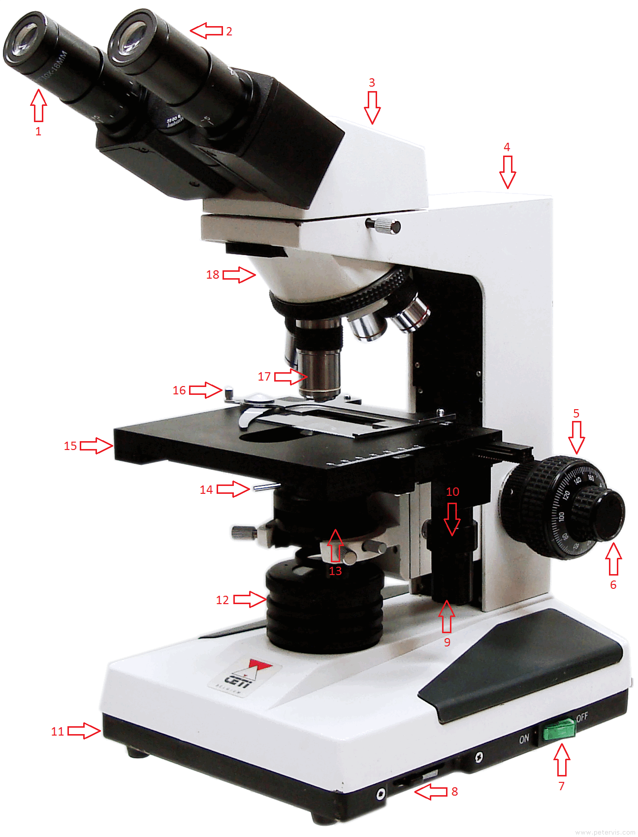
5. Knobs (fine and coarse) By adjusting the knob, you can adjust the focus of the microscope. The majority of the microscope models today have the knobs mounted on the same part of the device. Image 5: The circled parts of the microscope are the fine and coarse adjustment knobs. Picture Source: bp.blogspot.com.
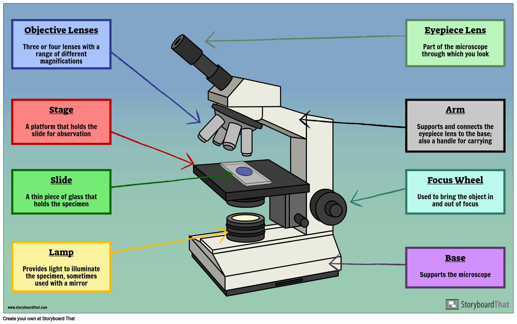
Labelled Microscope with Functions Storyboard by oliversmith
The description given below summarize the brief description of microscope parts used to visualize the microscopic specimens such as animal cells, plant cells, microbes, bacteria, viruses, microorganisms etc. The Microscopes parts divided into three different structural parts Head, Base, and Arms. Head/Body: It contain the optical parts in the.
