A Visual Guide to Dog Anatomy (Muscle, Organ & Skeletal Drawings) All Things Dogs

Unit 10 L3 Animation & Games Portfolio
Diaphragm: The diaphragm is the primary muscle involved in breathing. When a dog barks, it contracts the diaphragm forcefully to expel air out of its lungs and through its vocal cords. Laryngeal muscles: The laryngeal muscles control the opening and closing of the dog's vocal cords, which are located in the larynx (voice box) in the neck.

Anatomy Of Dog Spine Canis Lupus German Shepherd Skeleton Dog sketch, Dog anatomy, Dog skeleton
Bones - Dog Anatomy Great Dane and Chihuahua Skeletons, Sklmsta, March 2010 Contents 1 Introduction 2 Anatomy of the Head 2.1 Skull 2.1.1 Occipital Bone (os occipitale) 2.1.2 Sphenoid Bone (os sphenoidale) 2.1.3 Temporal Bone (os temporale) 2.1.4 Frontal Bone (os frontale) 2.1.5 Parietal Bone (os parietale) 2.1.6 Ethmoid Bone (os ethmoidale)

PetMassage Chart 3 Skeleton of the Dog · PetMassage™ Training and Research Institute
Dog anatomy comprises the anatomical studies of the visible parts of the body of a domestic dog.Details of structures vary tremendously from breed to breed, more than in any other animal species, wild or domesticated, as dogs are highly variable in height and weight. The smallest known adult dog was a Yorkshire Terrier that stood only 6.3 cm (2.5 in) at the shoulder, 9.5 cm (3.7 in) in length.

Dog Anatomy Hip Joints LOANKAS
They can hear sounds that are undetectable to the human ear. As compared to the 2 to 3 million scent glands that humans possess, dogs have between 200 to 300 million. The tail set is from where the tail begins. Some dogs have high-set tails, while some have low-set tails. Like the elbow on the foreleg, there is a hock present on the hind leg.

Anatomy of dog skeleton with labeled inner bone scheme vector illustration Dog skeleton
Labeled anatomy of the head and skull of the dog on CT imaging (bones of cranium, brain, face, paranasal sinus, muscles of head) This module of vet-Anatomy presents an atlas of the anatomy of the head of the dog on a CT. Images are available in 3 different planes (transverse, sagittal and dorsal), with two kind of contrast (bone and soft tissues).
Dog Skeletal Skull Anatomy Poster 18 X 24 Etsy
This veterinary anatomical atlas includes selected labeling structures to help student to understand and discover animal anatomy (skeleton, bones, muscles, joints, viscera, respiratory system, cardiovascular system). Positional and directional terms, general terminology and anatomical orientation are also illustrated.

How to Improve Dog Joint Health and Understand Joint Problems and Ortho Dog
Humans have three muscles whereas dogs have 18, and a dog can hear frequencies up to 65 kHz, while a human can only hear in the 12 to 20 kHz range. Dogs have about 320 bones, which can vary slightly depending on the presence of dewclaws and their tail structure. Humans have 206 bones.

FileDog anatomy lateral skeleton view.jpg
The scapula is a flat triangular bone at the top of the shoulder; more commonly known as the shoulder blade. It consists of 2 surfaces (medial and lateral), 3 borders (cranial, caudal and dorsal) and 3 angles (craniodorsal, caudodorsal and ventral angle).. Dog Anatomy project is developed by Veterinary Technician Program and The Sheridan.

Best Guardian Dog Rottweiler's Anatomy Dog anatomy, Rottweiler, Dog leg
ISSN 2534-5087. This veterinary anatomy module of the dog contains 218 illustrations dedicated to the canine osteology anatomy. Here are presented scientific illustrations of the canine skeleton, with the main dog's bones and its structures displayed from different anatomical standard views (cranial, caudal, lateral, medial, dorsal, palmar..).

Dog Anatomy
Anatomic Planes The main planes of motion for dogs are as follows (see Figure 5-1): • The sagittal plane divides the dog into right and left portions. If this plane were in the midline of the body, this is the median plane or median sagittal plane. • The dorsal plane divides the dog into ventral and dorsal portions.

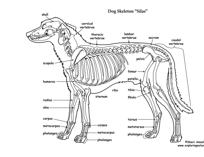
Dog skeleton 101 Dog Anatomy Bones Animal Hackers
Perhaps the most significant is head shape. There are three main different types of head formation in dogs: Dolichocephaly: dolichocephalic dogs are those with a head which is longer than it is wide. The skull and snout are elongated and their eyes are located in a lateral position, making it difficult to see well bifocally.

Dog skeleton with major bone elements labeled (Davis, 1987, p. 54;... Download Scientific Diagram
Pet Owner Version Components of the Musculoskeletal System in Dogs By Stephen B. Adams , DVM, DACVS, Department of Veterinary Clinical Sciences, College of Veterinary Medicine, Purdue University Reviewed/Revised Mar 2018 | Modified Oct 2022 Bones provide rigid structure to the body and shield internal organs from damage.
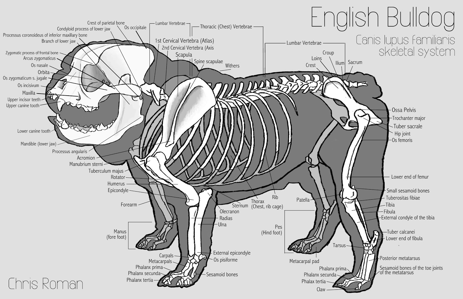
Chris Roman English Bulldog Anatomy Study
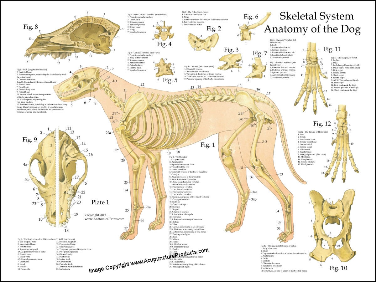
The dog skeleton is the bony part of dogs made for the support and protection of internal organs. Bones are connected through joints and muscles move the bones to produce the normal dog movements. In this article we will cover: Bone types and parts of the dog skeleton The dog skull Dog cranium The spine The Trunk The Forelimb The Hindlimb
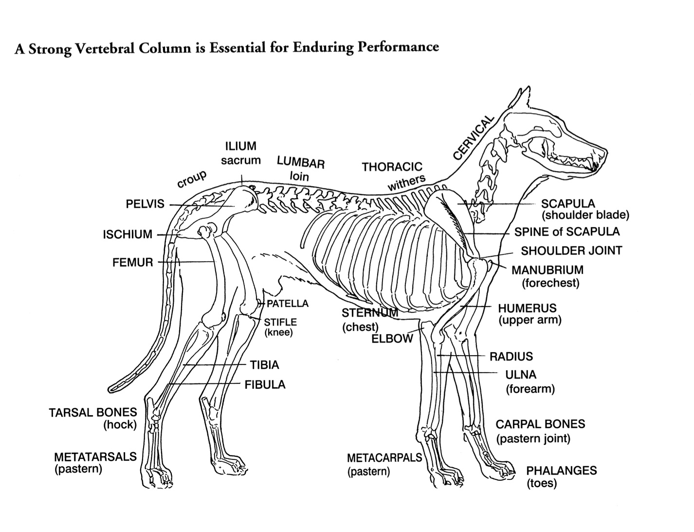
Helen King on Structure Evaluation Susan Garrett's Dog Training Blog
The Ultimate Guide to Dog Anatomy: Counting Dog Bones. Dogs, like humans and many other mammals, have a skeleton made up of bones that provide support, protection, and help with movement. The number of bones in a dog's body varies depending on its size and breed, but the average adult dog has about 319 bones. Puppies, on the other hand, have.
Dog Skeletal Anatomy
Summary Anatomy of a Dog Dog anatomy details the various structures of canines (e.g. muscle, organ and skeletal anatomy). The detailing of these structures changes based on dog breed due to the huge variation of size in dog breeds. Would you be surprised to know that short dogs are more aggressive? Or taller dogs are more affectionate?

Anatomy of a male dog crosssection, showing the skeleton and internal organs. Colour process
The anatomy of the temporal bone and the ear is complex as this region concentrates a large number of bony, muscular, articular, vascular and nervous structures. The purpose of the current anatomy module is to describe the normal anatomy of the inner and middle ear of the dog as depicted using CT of the temporal bone. Material and methods
