Biology Cell Structure and Functions
Biology Club Our cells 1 ( structure, function, division, disorder & cycle )
A cell is the smallest living thing in the human organism, and all living structures in the human body are made of cells. There are hundreds of different types of cells in the human body, which vary in shape (e.g. round, flat, long and thin, short and thick) and size (e.g. small granule cells of the cerebellum in the brain (4 micrometers), up to the huge oocytes (eggs) produced in the female.
Biology Cell Structure and Functions
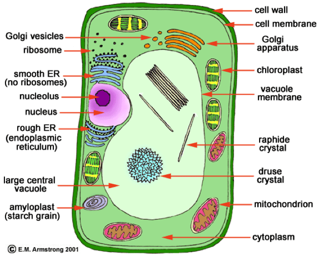
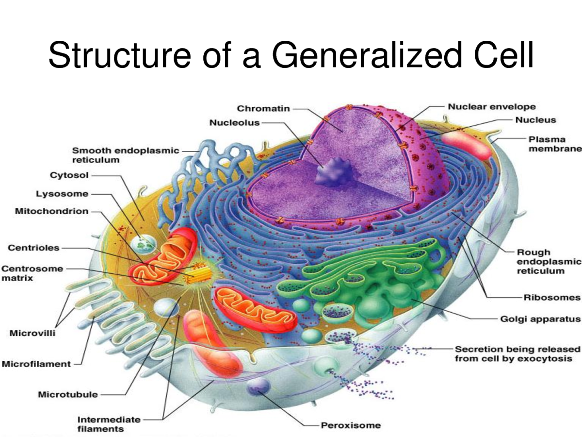
The plant cell wall is outside the cell membrane, and it provides structure for the cell. On the left is a circle representing an animal cell. The cell contains many cell parts with different shapes. A small bean-shaped cell part is labeled mitochondrion. A medium-sized circular cell part that has squiggly lines inside is labeled nucleus.
Eukaryotic cell structure diagrams Biological Science Picture Directory
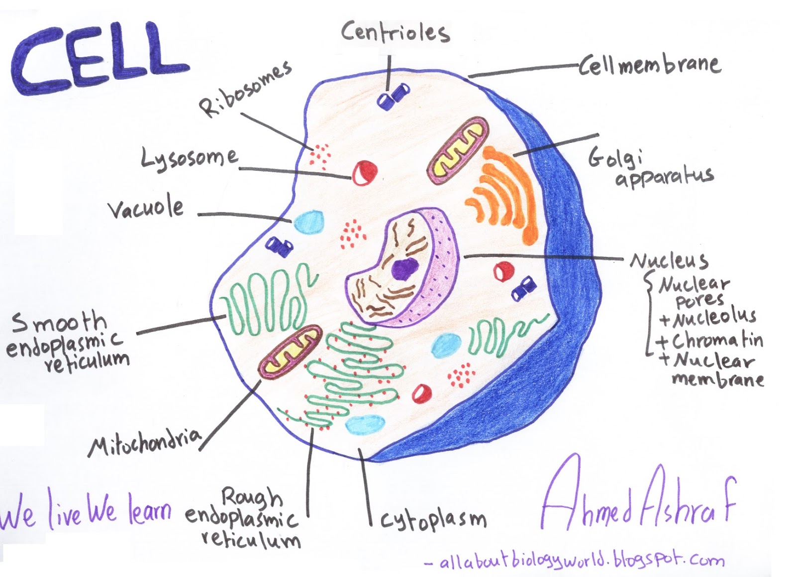
Cell diagram labeled. For this exercise we'll start with an image of a cell diagram ready labeled. Study this and make sure that you're clear about which structure is found where. Cell diagram unlabeled. It's time to label the cell yourself! As you fill in the cell structure worksheet, remember the functions of each part of the cell that.

animal cell diagram easy Kris Hammett
The cell structure illustrations for these diagrams were generated in BioRender. Both diagrams feature a drag-and-drop labelling activity created with H5P here on Learnful. These h5p resources are made available openly with the CC BY license. Plant Cell Structure: Animal Cell Structure:

Cells
Labeled Animal Cell Diagram. Blank Animal Cell Diagram Worksheet. The third and fourth diagrams are animal cell diagram worksheets. Quiz yourself by filling in the blanks. Unlabeled Animal Cell Diagram. Finally, an unlabeled version of the diagram is included at the bottom of the page, in color and black and white. This may be useful as a.
Structure of cell Cell structure and functions, Class 8
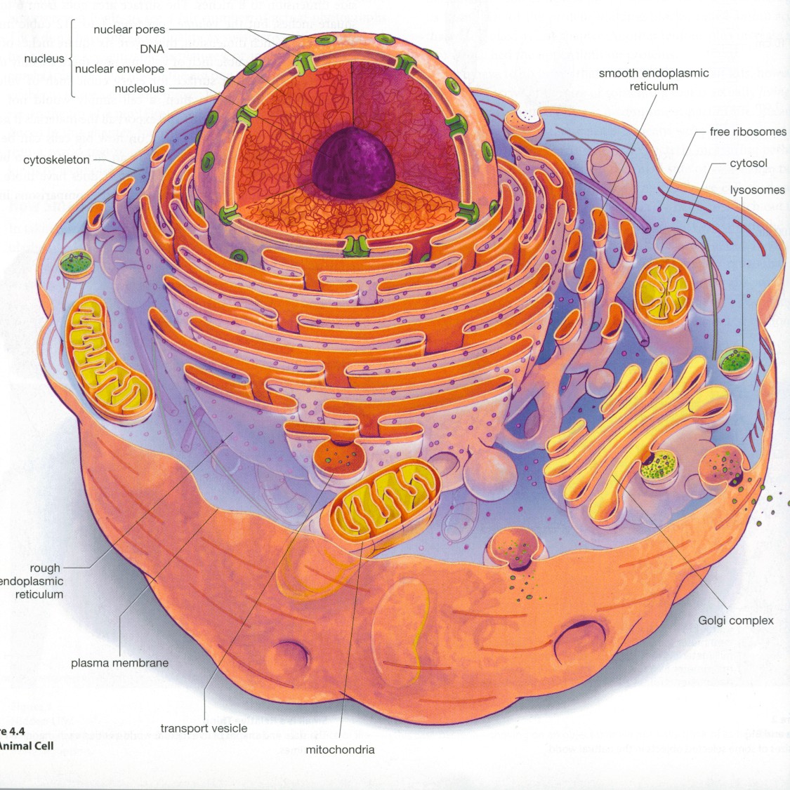
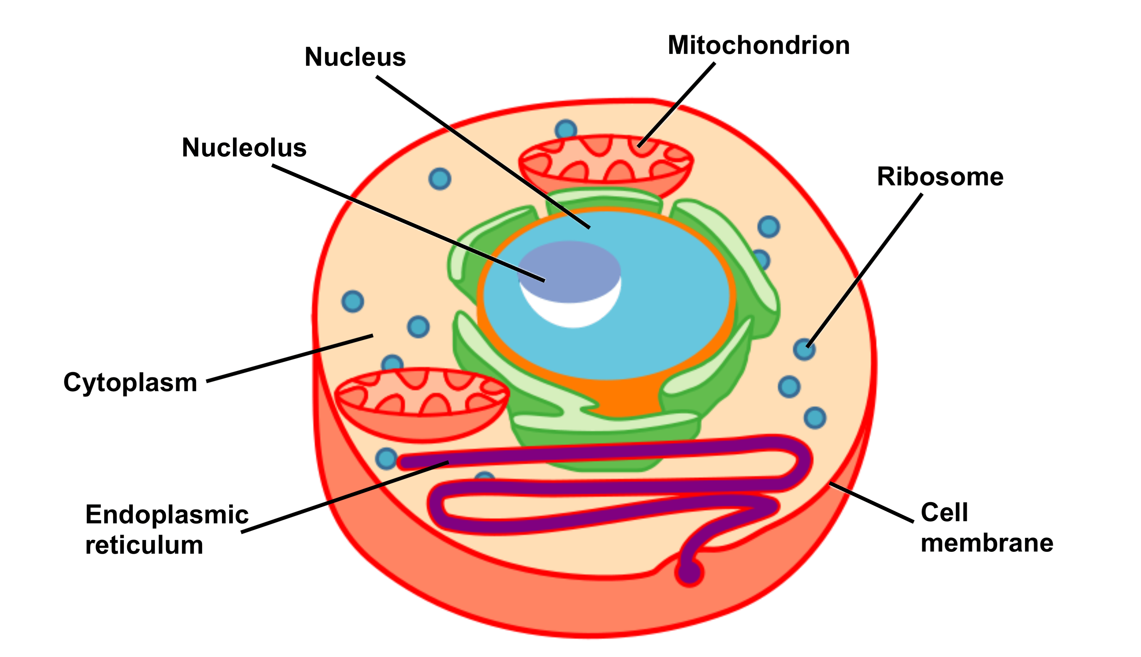
Labeled diagram of a typical animal cell Nucleus. The nucleus contains all the genetic material in a cell. This genetic information is called deoxyribonucleic acid (DNA). DNA contains all the instructions for making proteins, which control all of the body's activities. Therefore, the nucleus is like the manager's office of the cell.

Cell Structure
A plasma membrane encloses the cell contents of both plant and animal cells, but it is the outer coating of an animal cell. Animal Cell Structure: Organelles and Their Functions. Worksheet: Label the Parts of an Animal Cell [Google Apps worksheet][worksheet PDF][worksheet PNG][answers PNG] Common Questions About Animal Cells.

Animal Cell diagram with labels by Russell Kightley Media
Structure of a cell: Quiz 2; Structure of a cell: Unit test; About this unit. This unit is part of the Biology library. Browse videos, articles, and exercises by topic. Introduction to cells. Start your cellular journey the right way: with some history and some microscopy! Here, we'll learn more about how cells were discovered, how they can be.

South Pontotoc Biology Plant and Animal Cell Diagrams
Animal cell size and shape. Animal cells come in all kinds of shapes and sizes, with their size ranging from a few millimeters to micrometers. The largest animal cell is the ostrich egg which has a 5-inch diameter, weighing about 1.2-1.4 kg and the smallest animal cells are neurons of about 100 microns in diameter.

Biology 101 Cells Owlcation
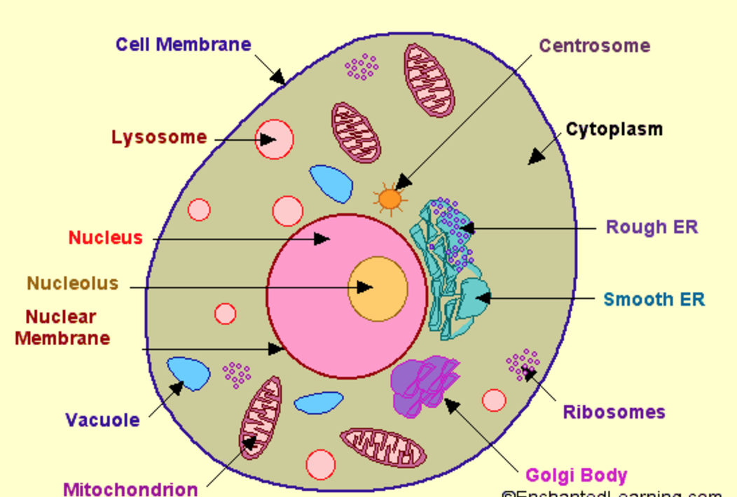
As observed in the labeled animal cell diagram, the cell membrane forms the confining factor of the cell, that is it envelopes the cell constituents together and gives the cell its shape, form, and existence. Cell membrane is made up of lipids and proteins and forms a barrier between the extracellular liquid bathing all cells on the exterior.
Animal Cell Well Labelled Diagram Preview We did not find results for
Diagram of a plant cell with components labeled. Image modified from OpenStax Biology. [Attribution and references]. is the transport network of the cell and it extends from and connects the nuclear membrane to the plasma membrane of a cell. But then whenever we draw a diagram of a typical plant or animal cell, we never extend it to the.

What is a cell? Facts
The animal cell diagram is widely asked in Class 10 and 12 examinations and is beneficial to understand the structure and functions of an animal. A brief explanation of the different parts of an animal cell along with a well-labelled diagram is mentioned below for reference. Also Read Different between Plant Cell and Animal Cell
Cell Structure and Function Part 1 The Organelles Medical Exam Prep
Cell labelling diagram. Below are some examples of what cells labelled with fluorescent dyes might look like. Hopefully, you now have more understanding of cell labelling and how useful it is! Cell Labeling - Key takeaways.

Plant Cell Labelled Diagram Ideas of Europedias
Animal cells have a basic structure. Below the basic structure is shown in the same animal cell, on the left viewed with the light microscope, and on the right with the transmission electron.

Organelles, cell growth and cytoskeleton Diagram Quizlet
Cell Notation (Cell Diagrams) Recall that standard cell potentials can be calculated from potentials E 0 cell for both oxidation and reduction reactions. A positive cell potential indicates that the reaction proceeds spontaneously in the direction in which the reaction is written. Conversely, a reaction with a negative cell potential proceeds.

Explain the nucleus of a cell with a neat labeled diagram Science Cell Structure and
Eukaryotic cells also have organelles, which are membrane-bound structures found within the cell. If you looked at eukaryotic cells under a microscope, you'd see distinct structures of all shapes and sizes. Prokaryotic cells, on the other hand, would look more uniform because they don't have those membrane-bound structures to break up the cell.
